Skip to Content
Merck
All Photos(6)

Key Documents

MABN92

Sigma-Aldrich

Anti-Iba1/AIF1 Antibody

clone 20A12.1, from mouse

Synonym(s):

Allograft inflammatory factor 1, AIF-1, Ionized calcium-binding adapter molecule 1, Protein G1

Sign Into View Organizational & Contract Pricing


About This Item

UNSPSC Code:
12352203
eCl@ss:
32160702
NACRES:
NA.41

biological source

mouse

Quality Level

antibody form

purified antibody

antibody product type

primary antibodies

clone

20A12.1, monoclonal

species reactivity

rat, mouse, human

packaging

antibody small pack of 25 μg

technique(s)

immunohistochemistry: suitable
western blot: suitable

isotype

IgG1κ

NCBI accession no.

UniProt accession no.

shipped in

ambient

storage temp.

2-8°C

target post-translational modification

unmodified

Gene Information

human ... AIF1(199)

General description

The allograft inflammatory factor-1 (AIF-1) is a 17 kDa interferon-gamma (IFN-gamma) inducible Ca(2+)-binding EF-hand protein that is encoded within the HLA class III genomic region. AIF-1 is known to be identical with ionized Ca(2+)-binding adaptor 1, microglia response factor-1, and daintain. AIF-1 expression is mostly limited to the macrophage/monocyte lineage, and is augmented by cytokines, such as IFN-gamma. It is reported to be involved in neuronal function, inflammatory responses, allograft rejection, autoimmunity, and the activation and function of macrophages. It is also shown to be expressed in macrophages and microglial cells in autominnume disesases such as experimental autoimmune encephalomyelitis, and neuritis and uveitis models.

Immunogen

Linear peptide corresponding to human Iba1/AIF1.

Application

Anti-Iba1/AIF1 Antibody is an antibody against Iba1/AIF1 for use in WB, IH.
Research Category
Neuroscience
Research Sub Category
Developmental Neuroscience
Western Blot Analysis: 1 µg/mL from a representative lot detected Iba1/AIF1 in 10 µg of mouse thymus tissue lysate.

Western Blot Analysis: 1 µg/mL from a representative lot detected Iba1/AIF1 in 10 µg of rat thymus tissue lysate.

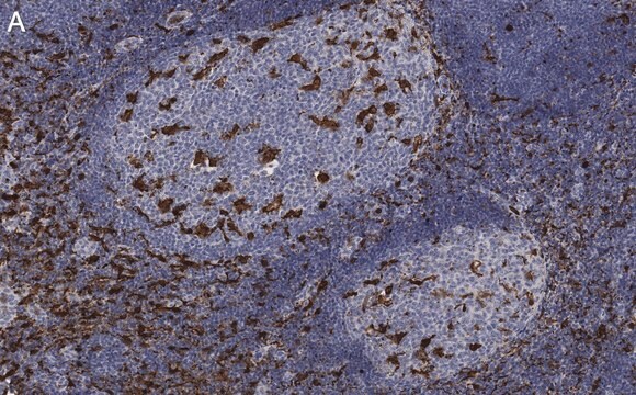
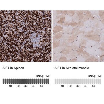
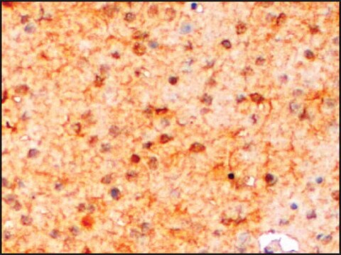
Immunohistochemistry Analysis: A 1:300 dilution from a representative lot detected Iba/AIF1 in human spleen tissue.
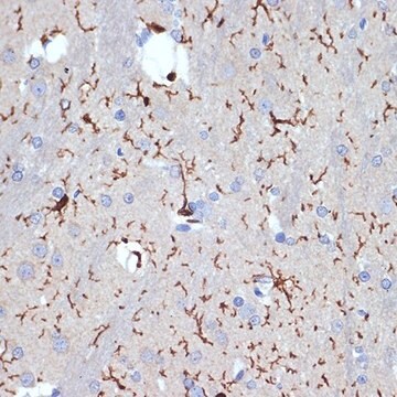
Immunohistochemistry Analysis: A 1:1000 dilution from a representative lot detected Iba/AIF1 in rat spleen tissue

Quality

Evaluated in Western Blot in human thymus tissue lysate.

Western Blot Analysis: 1 µg/mL of this antibody detected Iba1/AIF1 in 10 µg of human thymus tissue lysate.

Target description

~17 kDa observed

Physical form

Format: Purified
Protein G Purified
Purified mouse monoclonal IgG1κ in buffer containing 0.1 M Tris-Glycine (pH 7.4), 150 mM NaCl with 0.05% sodium azide.

Storage and Stability

Stable for 1 year at 2-8°C from date of receipt.

Analysis Note

Control
Human thymus tissue lysate

Other Notes

Concentration: Please refer to the Certificate of Analysis for the lot-specific concentration.

Disclaimer

Unless otherwise stated in our catalog or other company documentation accompanying the product(s), our products are intended for research use only and are not to be used for any other purpose, which includes but is not limited to, unauthorized commercial uses, in vitro diagnostic uses, ex vivo or in vivo therapeutic uses or any type of consumption or application to humans or animals.

Not finding the right product?  

Try our Product Selector Tool.

Storage Class Code

12 - Non Combustible Liquids

WGK

WGK 1

Flash Point(F)

Not applicable

Flash Point(C)

Not applicable


Certificates of Analysis (COA)

Search for Certificates of Analysis (COA) by entering the products Lot/Batch Number. Lot and Batch Numbers can be found on a product’s label following the words ‘Lot’ or ‘Batch’.

Already Own This Product?

Find documentation for the products that you have recently purchased in the Document Library.

Visit the Document Library

Rahmeh Othman et al.
Pharmaceuticals (Basel, Switzerland), 13(6) (2020-07-01)
Kinins are vasoactive peptides and mediators of inflammation, which signal through two G protein-coupled receptors, B1 and B2 receptors (B1R, B2R). Recent pre-clinical findings suggest a primary role for B1R in a rat model of wet age-related macular degeneration (AMD).
Macrophage Ablation Reduces M2-Like Populations and Jeopardizes Tumor Growth in a MAFIA-Based Glioma Model.
Gabrusiewicz, K; Hossain, MB; Cortes-Santiago, N; Fan, X; Kaminska, B; Marini, FC; Fueyo et al.
Neoplasia null
Chelsea M Larabee et al.
Molecular vision, 22, 332-341 (2016-04-29)
Optic neuritis affects most patients with multiple sclerosis (MS), and current treatments are unreliable. The purpose of this study was to characterize the contribution of Th1 and Th17 cells to the development of optic neuritis. Mice were passively transferred myelin-specific
Barbara D'Angelo et al.
Neuropharmacology, 101, 13-23 (2015-09-20)
Hypoxic-ischaemic (HI) injury is an important cause of neurological morbidity in neonates. HI leads to pathophysiological responses, including inflammation and oxidative stress that culminate in cell death. Activation of glycogen synthase kinase 3β (GSK3β) and the signal transducer and activator
Jenna M Ziebell et al.
Neuroscience, 359, 209-223 (2017-07-25)
Diffuse traumatic brain injury (TBI) initiates secondary pathology, including inflammation and reduced myelination. Considering these injury-related pathologies, the many states of activated microglia as demonstrated by differing morphologies would form, migrate, and function in and through fields of growth-inhibitory myelin

Our team of scientists has experience in all areas of research including Life Science, Material Science, Chemical Synthesis, Chromatography, Analytical and many others.

Contact Technical Service