Skip to Content
Merck
All Photos(3)

Key Documents

MABN1185

Sigma-Aldrich

Anti-Tau (4-repeat isoform RD4) Antibody, clone 7D12.1

clone 7D12.1, from mouse

Synonym(s):

Microtubule-associated protein tau, 4-repeat isoforms, Neurofibrillary tangle protein, 4-repeat isoforms, Paired helical filament-tau, 4-repeat isoforms, PHF-tau, PNS-tau, Tau-D, Tau-E, Tau-F, Tau-G

Sign Into View Organizational & Contract Pricing


About This Item

UNSPSC Code:
12352203
eCl@ss:
32160702
NACRES:
NA.41

biological source

mouse

Quality Level

antibody form

purified antibody

antibody product type

primary antibodies

clone

7D12.1, monoclonal

species reactivity

rat, human

species reactivity (predicted by homology)

mouse (based on 100% sequence homology), bovine (based on 100% sequence homology)

technique(s)

immunohistochemistry: suitable (paraffin)
western blot: suitable

isotype

IgG1κ

NCBI accession no.

UniProt accession no.

target post-translational modification

unmodified

Gene Information

General description

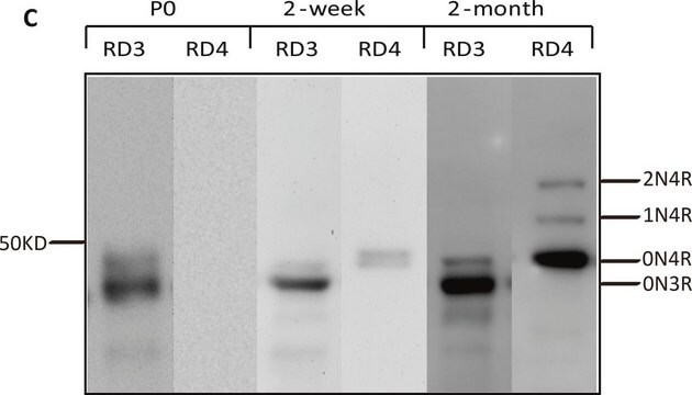
Microtubule-associated protein tau (UniProt P10636; also known as Neurofibrillary tangle protein, Paired helical filament-tau, PHF-tau) is encoded by the MAPT (also known as MAPTL, MTBT1, TAU) gene (Gene ID 4137) in human. Extracellular plaque deposits composed of amyloid-β and intracellular neurofibrillary tangles (NFTs) composed of truncated and hyperphosphorylated tau are two neuropathological hallmarks of Alzheimer′s disease (AD). Tau pathology (tauopathy) can cause toxicity when the brain is devoid of amyloid plaques, and tangle pathology correlates better with clinical dementia than amyloid pathology. Abnormal processing of tau by hyperphosphorylation and proteolytic truncation contribute to the toxicity of tau. Tau also undergoes other types of posttranslational modifications, including glycosylation, ubiquitination, glycation, polyamination, nitration, and lysine methylation, which are believed to be important for its non-pathological functions, including polymerization and stabilization of microtubules. Alternative splicings result in multiple Tau isoforms, including five human Tau spliced isoforms with four C-terminal microtubule-binding repeats/domains (RD4) and four spliced isoforms that contain only three microtubule-binding domains (RD3) due to the absence of exon 10-conding sequence. Isoform-specific antibodies are useful tools for analyzing tau isoforms expression and distribution as well as pathological changes in the human brain.

Specificity

Clone 7D12.1 targets an epitope sequence present in Tau spliced isoforms with four C-terminal microtubule-binding repeats/domains (RD4), including human isoforms PNS-tau, Tau-D, Tau-E, Tau-F, and Tau-G, but not Tau spliced isoforms that contain only three microtubule-binding domains (RD3) due to the absence of exon 10-conding sequence (human isoforms Fetal-tau, Tau-A, Tau-B, and Tau-C).

Immunogen

Epitope: Junction-flanking region coded by adjacent exons 9 & 10.
KLH-conjugated synthetic peptide (VQIINKKLDLSNVQSKC) corresponding to a junction-flanking sequence coded by adjacent exons 9 & 10 of Tau. The immunizing sequence is identical in human, rat, mouse and bovine.

Application

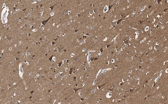
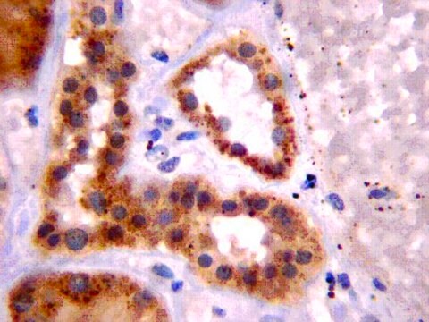
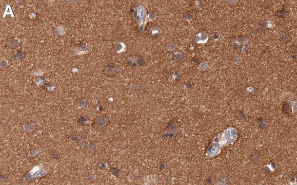
Immunohistochemistry Analysis: A 1:50-250 dilution from a representative lot detected Tau 4-repeat isoforms in Alzheimer′s diseased (AD) human brain, as well as human kidney and non-AD brain tissue sections.
Research Category
Neuroscience
Research Sub Category
Neurodegenerative Diseases
This Anti-Tau Antibody, clone 7D12.1 is validated for use in Western Blotting, Immunohistochemistry (Paraffin) for the detection of Tau (4-repeat isoform RD4).

Quality

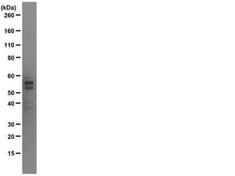
Evaluated by Western Blotting in rat brain tissue cytosol lysate.

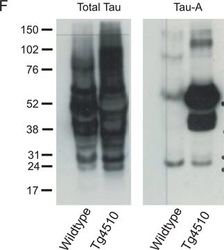
Western Blotting Analysis: 0.1 µg/mL of this antibody detected Tau 4-repeat isoforms in 10 µg of rat brain tissue cytosol lysate.

Target description

~45-58 kDa observed. 78,80 kDa (human isoform 1; PNS-tau), 39.88 kDa (human isoform 6; Tau-D), 42.84 kDa (human isoform 7; Tau-E), 45.72 kDa (human isoform 8; Tau-F), 80.81 kDa (human isoform 9; Tau-G) calculated (Met1 removed).

Physical form

Format: Purified
Protein G purified.
Purified mouse monoclonal IgG1κ antibody in buffer containing 0.1 M Tris-Glycine (pH 7.4), 150 mM NaCl with 0.05% sodium azide.

Storage and Stability

Stable for 1 year at 2-8°C from date of receipt.

Other Notes

Concentration: Please refer to lot specific datasheet.

Disclaimer

Unless otherwise stated in our catalog or other company documentation accompanying the product(s), our products are intended for research use only and are not to be used for any other purpose, which includes but is not limited to, unauthorized commercial uses, in vitro diagnostic uses, ex vivo or in vivo therapeutic uses or any type of consumption or application to humans or animals.

Not finding the right product?  

Try our Product Selector Tool.

Storage Class Code

12 - Non Combustible Liquids

WGK

WGK 1

Flash Point(F)

Not applicable

Flash Point(C)

Not applicable


Certificates of Analysis (COA)

Search for Certificates of Analysis (COA) by entering the products Lot/Batch Number. Lot and Batch Numbers can be found on a product’s label following the words ‘Lot’ or ‘Batch’.

Already Own This Product?

Find documentation for the products that you have recently purchased in the Document Library.

Visit the Document Library

Susan C Schwerin et al.
Journal of neuropathology and experimental neurology, 80(2), 112-128 (2021-01-10)
Blast exposures are a hallmark of contemporary military conflicts. We need improved preclinical models of blast traumatic brain injury for translation of pharmaceutical and therapeutic protocols. Compared with rodents, the ferret brain is larger, has substantial sulci, gyri, a higher

Our team of scientists has experience in all areas of research including Life Science, Material Science, Chemical Synthesis, Chromatography, Analytical and many others.

Contact Technical Service