Skip to Content
Merck
All Photos(1)

Documents

72485

Sigma-Aldrich

Nile Red

for microscopy

Synonym(s):

Nile Blue A Oxazone

Sign Into View Organizational & Contract Pricing


About This Item

Empirical Formula (Hill Notation):
C20H18N2O2
CAS Number:
Molecular Weight:
318.37
Beilstein:
279110
EC Number:
MDL number:
UNSPSC Code:
12171500
PubChem Substance ID:
NACRES:
NA.32

grade

for microscopy

form

crystals

mp

203-205 °C (lit.)

λmax

553 nm

SMILES string

CCN(CC)c1ccc2N=C3C(Oc2c1)=CC(=O)c4ccccc34

InChI

1S/C20H18N2O2/c1-3-22(4-2)13-9-10-16-18(11-13)24-19-12-17(23)14-7-5-6-8-15(14)20(19)21-16/h5-12H,3-4H2,1-2H3

InChI key

VOFUROIFQGPCGE-UHFFFAOYSA-N

Looking for similar products? Visit Product Comparison Guide

General description

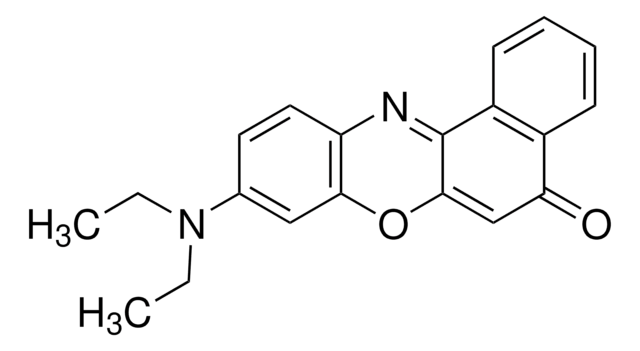
Nile Red is a phenoxazone dye. It is an important stain, which is used to detect intracellular lipid droplets and proteins. Nile Red detects lysosomes and lysosome-related organelles, like gut granules in C. elegans intestinal cells. It exhibits strong fluorescence enhancement and is used for staining SDS (sodium dodecyl sulphate) gels.

Application

Nile Red has been used:
  • as a fluorescent dye for staining fat by confocal scanning laser microscopy (CSLM)
  • to label nanoparticles (NPs)
  • for lipid analysis

Storage Class Code

11 - Combustible Solids

WGK

WGK 3

Flash Point(F)

Not applicable

Flash Point(C)

Not applicable

Personal Protective Equipment

dust mask type N95 (US), Eyeshields, Gloves

Certificates of Analysis (COA)

Search for Certificates of Analysis (COA) by entering the products Lot/Batch Number. Lot and Batch Numbers can be found on a product’s label following the words ‘Lot’ or ‘Batch’.

Already Own This Product?

Find documentation for the products that you have recently purchased in the Document Library.

Visit the Document Library

Influence of process parameters on formation of fibrous materials from dense calcium caseinate dispersions and fat
Manski JM, et al.
Food Hydrocolloids, 22(4), 587-600 (2008)
Labeling nanoparticles: Dye leakage and altered cellular uptake
Snipstad S, et al.
Cytometry. Part A : the Journal of the International Society For Analytical Cytology, 91(8), 760-766 (2017)
Protein Gel Staining Methods: An Introduction and Overview
Steinberg TH
Methods in Enzymology, 463, 541-563 (2009)
Importance of intrinsic properties of dense caseinate dispersions for structure formation
Manski JM, et al.
Biomacromolecules, 8(11), 3540-3547 (2007)
Analysis of membrane-bound organelles
Fares H and van der Bliek AM
Methods in Cell Biology, 107, 239-263 (2012)

Our team of scientists has experience in all areas of research including Life Science, Material Science, Chemical Synthesis, Chromatography, Analytical and many others.

Contact Technical Service