
推薦產品


生物源
rabbit
品質等級
重組細胞
expressed in HEK 293 cells
共軛
unconjugated
抗體表格
purified antibody
抗體產品種類
primary antibodies
無性繁殖
2E20, recombinant monoclonal
描述
2E20 Clone
產品線
ZooMAb® learn more
形狀
lyophilized
分子量
calculated mol wt 15.33 kDa
observed mol wt ~17 kDa
純化經由
using Protein A
物種活性
human
包裝
antibody small pack of 25 μL
環保替代產品特色
Waste Prevention
Designing Safer Chemicals
Design for Energy Efficiency
Learn more about the Principles of Green Chemistry.
加強驗證
recombinant expression
Learn more about Antibody Enhanced Validation
技術
affinity binding assay: suitable
immunohistochemistry: suitable
western blot: suitable
同型
IgG
表位序列
N-terminal half
Protein ID登錄號
UniProt登錄號
環保替代類別
運輸包裝
ambient
儲存溫度
2-8°C
基因資訊
human ... H3-3A(3020)
一般說明
We are committed to bringing you greener alternative products, which adhere to one or more of The 12 Principles of Green Chemistry. This antibody is Preservative-free, produced without the harm or sacrifice of animals and exceptionally stable to allow for ambient shipping and storage if needed and thus aligns with "Waste Prevention", "Designing Safer Chemicals" and "Design for Energy Efficiency". Click here for more information.
ZooMAb® antibodies represent an entirely new generation of recombinant monoclonal antibodies. Each ZooMAb® antibody is manufactured using our proprietary recombinant expression system, purified to homogeneity, and precisely dispensed to produce robust and highly reproducible lot-to-lot consistency. Only top-performing clones are released for use by researchers. Each antibody is validated for high specificity and affinity across multiple applications, including its most commonly used application. ZooMAb® antibodies are reliably available and ready to ship when you need them.
特異性
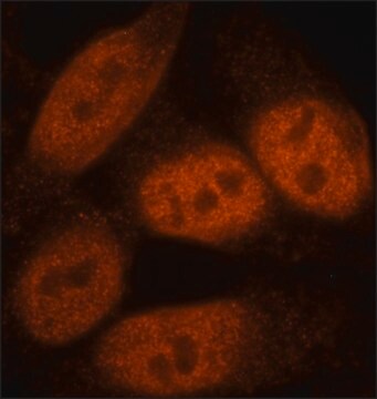
Clone 2E20 is a ZooMAb® rabbit recombinant monoclonal antibody that specifically detects Histone H3.3 with G34W mutation.
免疫原
KLH-conjugated linear peptide corresponding to 8 amino acids surrounding the mutated G34W site from the N-terminal half of human Histone H3.3.
應用
Quality Control Testing
Evaluated by Western Blotting in lysates from HEK293 cells expressing Histone H3.3 with G34W mutation.
Western Blotting Analysis (WB): A 1:10,000 dilution of this antibody detected Histone H3.3 G34W in lysate from HEK293 cells expressing this mutation, but not in wild-type HEK293 cells.
Tested Applications
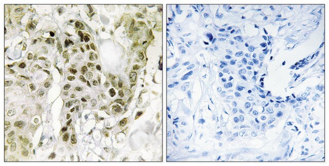
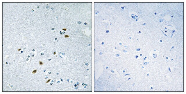
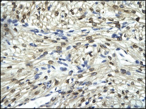
Immunohistochemistry (Paraffin) Analysis: A 1:100 dilution from a representative lot detected Histone H3.3 G34W in Human bone giant cell tumor tissue sections.
Affinity Binding Assay: A representative lot of this antibody bound Histone H3.3 G34W peptide with a KD of 3.1 x 10⁻⁶ in an affinity binding assay.
Note: Actual optimal working dilutions must be determined by end user as specimens, and experimental conditions may vary with the end user.
Evaluated by Western Blotting in lysates from HEK293 cells expressing Histone H3.3 with G34W mutation.
Western Blotting Analysis (WB): A 1:10,000 dilution of this antibody detected Histone H3.3 G34W in lysate from HEK293 cells expressing this mutation, but not in wild-type HEK293 cells.
Tested Applications
Immunohistochemistry (Paraffin) Analysis: A 1:100 dilution from a representative lot detected Histone H3.3 G34W in Human bone giant cell tumor tissue sections.
Affinity Binding Assay: A representative lot of this antibody bound Histone H3.3 G34W peptide with a KD of 3.1 x 10⁻⁶ in an affinity binding assay.
Note: Actual optimal working dilutions must be determined by end user as specimens, and experimental conditions may vary with the end user.
標靶描述
Histone H3.3 (UniProt: P84243; also known as H3.3) is encoded by the H3-3A (also known as H3.3A, H3F3, H3F3A, PP781, H3-3B) gene (Gene ID: 3020) in human. Histone H3 has two main variants, H3.1 and H3.3, which show different genomic localization patterns in animals. Histone H3.1 serves as the canonical histone, which is incorporated during DNA replication. Histone H3.3 is a highly conserved variant form of Histone H3, which replaces conventional H3 in a wide range of nucleosomes in active genes. Histone H 3.3 constitutes the predominant form of histone H3 in non-dividing cells and is incorporated into chromatin independently of DNA synthesis. H3.3 is associated with actively expressed genes in both animals and plants. It is predominantly enriched near transcription end sites (TES) of genes and positively associated with transcription. Histone H3 contains a main globular domain and a long N-terminal tail and is involved with the structure of the nucleosomes of the ′beads on a string′ structure. The N-terminal tail of histone H3 protrudes from the globular nucleosome core and can undergo several different types of epigenetic modifications that influence cellular processes. These modifications include the covalent attachment of methyl or acetyl groups to lysine and arginine amino acids and the phosphorylation of serine or threonine. For example, acetylation on Lys10 (H3K9ac; initial methionine removed) impairs methylation at Arg9 (H3R8me2s) and acetylation on Lys19 (H3K18ac) and Lys24 (H3K24ac) favors methylation at Arg18 (H3R17me). Phosphorylation at Ser11 (H3S10ph) by Aurora kinase B is crucial for chromosome condensation and cell-cycle progression during mitosis and meiosis. Phosphorylation at Ser11 by RPS6KA4 and RPS6KA5 is important during interphase because it enables the transcription of genes following external stimulation, like mitogens, stress, growth factors or UV irradiation and result in the activation of genes, such as c-Fos and c-Jun. Mutations in Histone H3.3 have been implicated in a high proportion of malignant pediatric brain cancers. The mutant H3.3 histone disrupts epigenetic post-translational modifications near genes involved in cancer processes and in brain function. Glycine 34 to tryptophan (G34W) mutation in Histone 3.3 have been linked to the development of giant cell tumor of bone (GCTB) and also in a mosaic disorder characterized by pheochromocytomas and paragangliomas. This ZooMAb® recombinant monoclonal antibody, generated by our propriety technology, offers significantly enhanced specificity, affinity, reproducibility, and stability over conventional monoclonals. (Ref.: Amary, F., et al. (2017). Am. J. Surg. Pathol. 41(8); 1059-1068).
外觀
Purified recombinant rabbit monoclonal antibody IgG, lyophilized in PBS with 5% Trehalose, normal appearance a coarse or translucent resin. The PBS/trehalose components in the ZooMAb formulation can have the appearance of a semi-solid (bead like gel) after lyophilization. This is a normal phenomenon. Please follow the recommended reconstitution procedure in the data sheet to dissolve the semi-solid, bead-like, gel-appearing material. The resulting antibody solution is completely stable and functional as proven by full functional testing. Contains no biocide or preservatives, such as azide, or any animal by-products. Larger pack sizes provided as multiples of 25 µL.
儲存和穩定性
Recommend storage of lyophilized product at 2-8°C; Before reconstitution, micro-centrifuge vials briefly to spin down material to bottom of the vial; Reconstitute each vial by adding 25 µL of filtered lab grade water or PBS; Reconstituted antibodies can be stored at 2-8°C, or -20°C for long term storage. Avoid repeated freeze-thaws.
其他說明
Concentration: Please refer to the Certificate of Analysis for the lot-specific concentration.
法律資訊
ZooMAb is a registered trademark of Merck KGaA, Darmstadt, Germany
免責聲明
Unless otherwise stated in our catalog or other company documentation accompanying the product(s), our products are intended for research use only and are not to be used for any other purpose, which includes but is not limited to, unauthorized commercial uses, in vitro diagnostic uses, ex vivo or in vivo therapeutic uses or any type of consumption or application to humans or animals.
未找到適合的產品?
試用我們的產品選擇工具.
儲存類別代碼
11 - Combustible Solids
水污染物質分類(WGK)
WGK 1
閃點(°F)
Not applicable
閃點(°C)
Not applicable
我們的科學家團隊在所有研究領域都有豐富的經驗,包括生命科學、材料科學、化學合成、色譜、分析等.
聯絡技術服務