推薦產品
材料
spherical
無菌
sterile; autoclaved
特點
lid: no
honeycomb diam. 3.4 mm
包裝
pack of 6 ea
製造商/商標名
MicroTissues Inc. 24-H
容積
20 μL
相關類別
一般說明
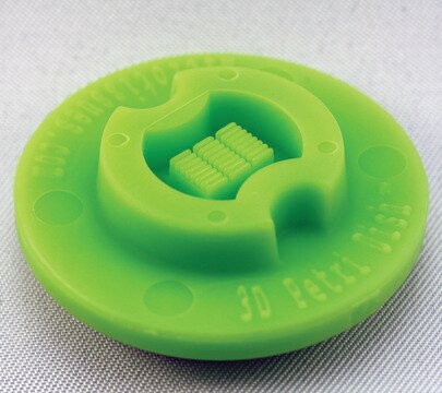
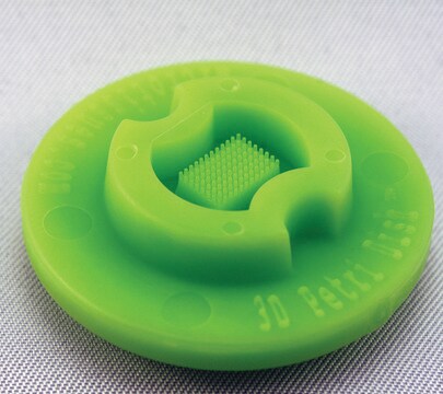
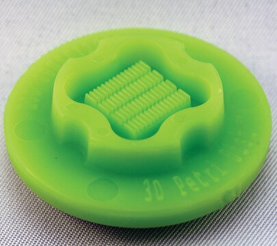
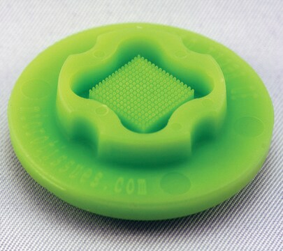
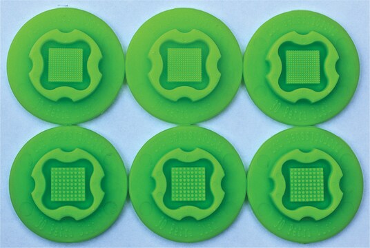
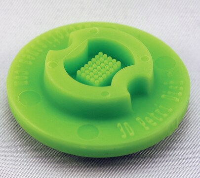
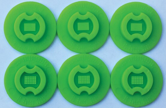
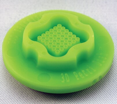
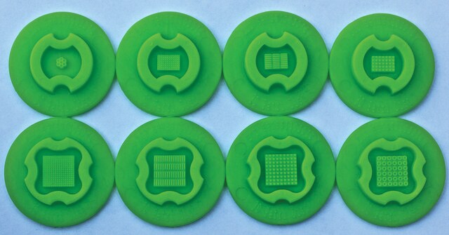
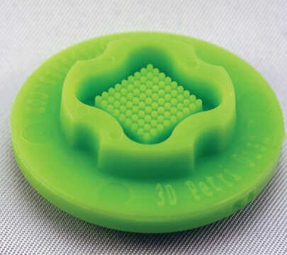
Six autoclavable precision micro-molds to cast 3D Petri Dish for forming honeycomb shaped microtissue. 3D Petri Dish for use in 24-well plate. Each micro-mold forms 1 honeycomb shaped recess.
When the gelled agarose is removed from the micro-mold, it is transferred to a standard 12 well or 24 well tissue culture dish and equilibrated with cell culture medium. Since the agarose is transparent, the spheroids or microtissues that form at the bottom of each agarose micro-well can be easily viewed using a standard inverted microscope using phase contrast, bright field or fluorescence microscopy.
- Nominal dimensions of each 3D culture recess: maximum size 3.4 mm
- Peg diam. 600 μm
- Trough W 400 μm x D 700 μm
- Micro-molds also form single chamber for cell seeding
When the gelled agarose is removed from the micro-mold, it is transferred to a standard 12 well or 24 well tissue culture dish and equilibrated with cell culture medium. Since the agarose is transparent, the spheroids or microtissues that form at the bottom of each agarose micro-well can be easily viewed using a standard inverted microscope using phase contrast, bright field or fluorescence microscopy.
- The micro-molds are reusable up to 12 times and can be sterilized via a standard steam autoclave (30 min, dry cycle)
- The micro-molds should be stored in a covered container to maintain sterility and to avoid collecting dust or fibres on their small features
- Any microscope with sufficient magnification and resolution functions for cell based views or images will be suitable for use with the 3D Petri Dish products
法律資訊
3D Petri Dish is a registered trademark of MicroTissues Inc.
MicroTissues is a registered trademark of MicroTissues Inc.
分析證明 (COA)
輸入產品批次/批號來搜索 分析證明 (COA)。在產品’s標籤上找到批次和批號,寫有 ‘Lot’或‘Batch’.。
客戶也查看了
B R Desroches et al.
American journal of physiology. Heart and circulatory physiology, 302(10), H2031-H2042 (2012-03-20)
To bridge the gap between two-dimensional cell culture and tissue, various three-dimensional (3-D) cell culture approaches have been developed for the investigation of cardiac myocytes (CMs) and cardiac fibroblasts (CFs). However, several limitations still exist. This study was designed to
我們的科學家團隊在所有研究領域都有豐富的經驗,包括生命科學、材料科學、化學合成、色譜、分析等.
聯絡技術服務