跳轉至內容
Merck
全部照片(1)

重要文件

C4585

Sigma-Aldrich

抗 β 微管蛋白 − 小鼠单克隆 Cy3 抗体 小鼠抗

clone TUB 2.1, purified from hybridoma cell culture

同義詞:

单克隆抗 β-微管蛋白

登入查看組織和合約定價


About This Item

分類程式碼代碼:
12352203
NACRES:
NA.41

生物源

mouse

品質等級

共軛

CY3 conjugate

抗體表格

purified from hybridoma cell culture

抗體產品種類

primary antibodies

無性繁殖

TUB 2.1, monoclonal

形狀

buffered aqueous solution

分子量

antigen 55 kDa

物種活性

human, rat, frog, moth, mouse, plant, rabbit, chicken, bovine, wheat, sea urchin, hamster

儲存條件

protect from light

技術

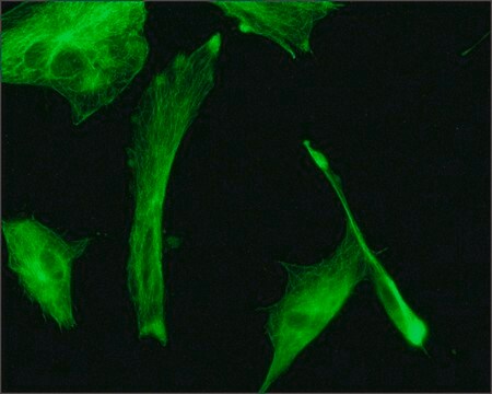
direct immunofluorescence: 1:100 using cultured chicken fibroblasts or BHK cells

同型

IgG1

運輸包裝

wet ice

儲存溫度

2-8°C

目標翻譯後修改

unmodified

尋找類似的產品? 前往 產品比較指南

一般說明

单克隆抗b微管蛋白(小鼠IgG1同种型)来源于小鼠骨髓瘤细胞和以纯化大鼠脑微管蛋白免疫的小鼠脾细胞融合产生的TUB2.1杂交瘤。微管蛋白是微管的主要成分。这种细胞内圆柱形丝状结构存在于几乎所有真核细胞中。微管蛋白是一种异二聚体,由α-微管蛋白和β-微管蛋白组成。这两个亚基的分子量均为55kDa,并具有可观的同源性。β-微管蛋白的结构特征在于具有被β-螺旋包围的两个α-折叠的核心。

特異性

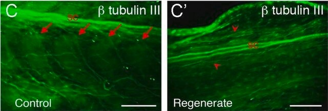
该抗体可识别 β 的所有 5 种亚型-微管蛋白(β1-β5).它在免疫印迹中与β-微管蛋白的羧基末端部分中的β-Lc和β-Sc片段反应。该抗体可用于定位培养细胞或组织切片中的 β-微管蛋白。

免疫原

来自大鼠脑的微管蛋白。

應用

小鼠源抗β-微管蛋白-Cy3抗体可用于:
  • 免疫组织化学分析
  • 免疫荧光
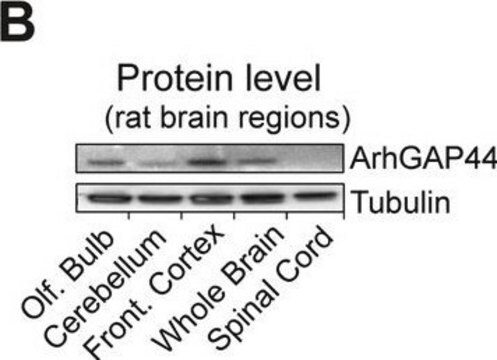
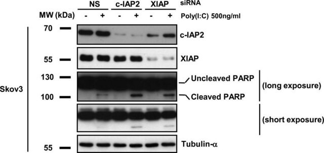
  • 蛋白质印迹
  • 免疫细胞化学
  • 免疫印迹
  • 免疫染色

生化/生理作用

微管在有丝分裂、细胞内转运、鞭毛运动和细胞骨架中起结构和移动元件的作用。β-微管蛋白基因突变可导致各种神经元迁移障碍,如无脑回、巨脑回和多小脑回畸形。

外觀

溶于0.01M磷酸盐缓冲盐水,pH 7.4,含有1%牛血清白蛋白以及15 mM叠氮化钠。

法律資訊

Cy由Amersham Biosciences Limited许可分销。

免責聲明

除非我们的产品目录或产品附带的其他公司文档另有说明,否则我们的产品仅供研究使用,不得用于任何其他目的,包括但不限于未经授权的商业用途、体外诊断用途、离体或体内治疗用途或任何类型的消费或应用于人类或动物。

未找到適合的產品?  

試用我們的產品選擇工具.

儲存類別代碼

10 - Combustible liquids

水污染物質分類(WGK)

nwg

閃點(°F)

Not applicable

閃點(°C)

Not applicable

個人防護裝備

Eyeshields, Gloves, multi-purpose combination respirator cartridge (US)


從最近期的版本中選擇一個:

分析證明 (COA)

Lot/Batch Number

未看到正確版本?

如果您需要一個特定的版本,您可以透過批號來尋找特定憑證。

已經擁有該產品?

您可以在文件庫中找到最近購買的產品相關文件。

存取文件庫

Liang Qiang et al.
Molecular biology of the cell, 21(2), 334-344 (2009-11-27)
The formation of interstitial axonal branches involves the severing of microtubules at sites where new branches form. Here we wished to ascertain whether basic fibroblast growth factor (bFGF) enhances axonal branching through alterations in proteins involved in the severing of
The microtubule-severing proteins spastin and katanin participate differently in the formation of axonal branches
Yu W, et al.
Molecular Biology of the Cell, 19(4), 1485-1498 (2008)
TAPping into the treasures of tubulin using novel protein production methods
Yu N and Galjart N
Essays in Biochemistry, 62(6), 781-792 (2018)
M Janmaleki et al.
Scientific reports, 6, 32418-32418 (2016-09-02)
This study focused on the effects of simulated microgravity (s-μg) on mechanical properties, major cytoskeleton biopolymers, and morphology of endothelial cells (ECs). The structural and functional integrity of ECs are vital to regulate vascular homeostasis and prevent atherosclerosis. Furthermore, these
Comprehensive genotype-phenotype correlation in lissencephaly
Tan AP, et al.
Quantitative imaging in medicine and surgery, 8(7), 673-673 (2018)

文章

Microtubules of the eukaryotic cytoskeleton are composed of a heterodimer of α- and β-tubulin. In addition to α-and β-tubulin, several other tubulins have been identified, bringing the number of distinct tubulin classes to seven.

我們的科學家團隊在所有研究領域都有豐富的經驗,包括生命科學、材料科學、化學合成、色譜、分析等.

聯絡技術服務