跳轉至內容
Merck
全部照片(4)

文件

PICM03050

Millipore

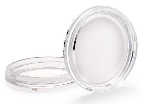
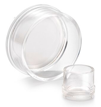
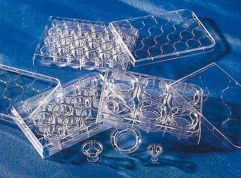
Millicell® Standing细胞培养池

pore size 0.4 μm, diam. 30 mm, transparent PTFE membrane, hydrophilic, H 13 mm, size 6 wells, sterile

同義詞:

Millicell细胞培养小室,30mm,亲水PTFE,0.4µm, 可渗透培养小室, 组织培养小室, 组织培养板, 细胞培养小室

登入查看組織和合約定價


About This Item

分類程式碼代碼:
41104923
eCl@ss:
32011202
NACRES:
NB.22

材料

polystyrene
transparent PTFE membrane

品質等級

無菌

ethylene oxide treated
sterile

特點

hydrophilic

包裝

pack of 50

製造商/商標名

Millicell®

參數

50 °C max. temp.

技術

cell attachment: suitable
cell culture | mammalian: suitable
cell differentiation: suitable

高度

13 mm

直徑

30 mm

過濾面積

4.2 cm2

尺寸

6 wells

表面積

4.2 cm2

有效容積

1.5 mL

顏色

transparent, when wetted

基質

Biopore

孔徑

0.4 μm pore size

結合類型

low binding surface

檢測方法

fluorometric

運輸包裝

ambient

一般說明

设备配置:6孔板,0.4μm孔径

使用Millicell®嵌套板,附着或悬浮细胞可以从其上下两侧接触到培养基。更加密切地模拟了体内所发生的细胞生长、结构和功能。另外,Millicell® 嵌套板使研究细胞单层的两侧成为可能。

Millicell® 立式立式嵌套板:
-特别有助于细胞成长,为细胞研究提供了绝佳机会

膜类型:
生物孔膜(亲水性 PTFE)
-用于低蛋白结合、活细胞观察和免疫荧光应用

应用
细胞附着、细胞生长、细胞分化、免疫细胞化学

應用

亲水性PTFE细胞培养嵌套板,孔径为0.4 µm,用于6孔培养板,适用于细胞附着、细胞培养、细胞分化、荧光 & 低蛋白结合。
研究类别
细胞培养

包裝

气泡袋独立包装

法律資訊

BIOPORE is a trademark of Merck KGaA, Darmstadt, Germany
Millicell is a registered trademark of Merck KGaA, Darmstadt, Germany

分析證明 (COA)

輸入產品批次/批號來搜索 分析證明 (COA)。在產品’s標籤上找到批次和批號,寫有 ‘Lot’或‘Batch’.。

已經擁有該產品?

您可以在文件庫中找到最近購買的產品相關文件。

存取文件庫

CB1 receptors and post-ischemic brain damage: Studies on the toxic and neuroprotective effects of cannabinoids in rat organotypic hippocampal slices.
Landucci, Elisa, et al.
Neuropharmacology, 60, 674-682 (2011)
Synapse elimination in olivo-cerebellar explants occurs during a critical period and leaves an indelible trace in Purkinje cells.
Letellier, Mathieu, et al.
Proceedings of the National Academy of Sciences of the USA, 106, 14102-14107 (2009)
Daniela Buonvicino et al.
British journal of pharmacology, 177(14), 3342-3356 (2020-03-22)
Drugs able to counteract progressive multiple sclerosis (MS) represent a largely unmet therapeutic need. Even though the pathogenesis of disease evolution is still obscure, accumulating evidence indicates that mitochondrial dysfunction plays a causative role in neurodegeneration and axonopathy in progressive
Preparation of organotypic hippocampal slice cultures: interface method.
De Simoni, Anna and Yu, Lily M Y
Nature Protocols, 1, 1439-1445 (2006)
Andrea S Cupp et al.
Biology of reproduction, 68(6), 2033-2037 (2003-02-28)
The first morphological event after initiation of male sex determination is seminiferous cord formation in the embryonic testis. Cord formation requires migration of pre-peritubular myoid cells from the adjacent mesonephros. The embryonic Sertoli cells are the first testicular cells to

文章

This page covers the basic indirect co-culture procedure on both sides of Millicell cell culture insert membranes.

條款

This protocol covers three methods for the microscopic examination of cell samples.

This protocol covers 3 modes for the microscopic examination of cell samples.

This page covers the indirect co-culture of embryonic stem cells with embryonic fibroblasts.

This page covers the basic indirect co-culture procedure on both sides of Millicell cell culture insert membranes.

相關內容

This page covers the ECM coating protocols developed for four types of ECMs on Millicell®-CM inserts, Collagen Type 1, Fibronectin, Laminin, and Matrigel.

This page covers the ECM coating protocols developed for four types of ECMs on Millicell®-CM inserts, Collagen Type 1, Fibronectin, Laminin, and Matrigel.

我們的科學家團隊在所有研究領域都有豐富的經驗,包括生命科學、材料科學、化學合成、色譜、分析等.

聯絡技術服務