APT428
CaspaTag Caspase 8 In Situ Assay Kit 25, Fluorescein
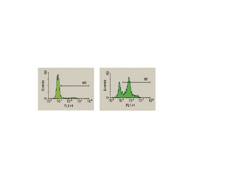
The CaspaTag Caspase-8 In Situ Assay Kit, Fluorescein for flow cytometry is a fluorescent-based assay for detection of active caspase-8 in cells undergoing apoptosis.
登入查看組織和合約定價
全部照片(1)
About This Item
分類程式碼代碼:
12161503
eCl@ss:
32161000
NACRES:
NA.84
推薦產品
品質等級
物種活性(以同源性預測)
mammals
製造商/商標名
CaspaTag
Chemicon®
技術
activity assay: suitable
flow cytometry: suitable
UniProt登錄號
檢測方法
fluorometric
運輸包裝
wet ice
一般說明
Apoptosis is an evolutionarily conserved form of cell suicide, which follows a specialized cellular process. The central component of this process is a cascade of proteolytic enzymes called caspases. These enzymes participate in a series of reactions that are triggered in response to pro-apoptotic signals and result in the cleavage of protein substrates, causing the disassembly of the cell1.
Caspases have been identified in organisms ranging from C. elegans to humans. The mammalian caspases play distinct roles in apoptosis and inflammation. In apoptosis, caspases are responsible for proteolytic cleavages that lead to cell disassembly (effector caspases), and are involved in upstream regulatory events (initiator caspases). An active caspase consists of two large and two small subunits that form two heterodimers, which associate in a tetramer2-4. In common with other proteases, caspases are synthesized as precursors that undergo proteolytic maturation, either autocatalytically or in a cascade by enzymes with similar specificity5.
Caspase enzymes specifically recognize a 4 or 5 amino acid sequence on the target substrate, which necessarily includes an aspartic acid residue. This residue is the target for cleavage, which occurs at the carbonyl end of the aspartic acid residue6. Caspases can be detected via immunoprecipitation, immunoblotting techniques using caspase specific antibodies, or by employing fluorochrome substrates, which become fluorescent upon cleavage by the caspase.
應用
Research Category
Apoptosis & Cancer
Apoptosis & Cancer
The CHEMICON® CaspaTag Caspase-8 In Situ Assay Kit, Fluorescein is a fluorescent-based assay for detection of active caspase-8 in cells undergoing apoptosis. The kit is for research use only. Not for use in diagnostic or therapeutic procedures.
Test Principle
CHEMICON′s CaspaTag Caspase-8 In Situ Assay Kits use a novel approach to detect active caspases. The methodology is based on Fluorochrome Inhibitors of Caspases (FLICA). The inhibitors are cell permeable and non-cytotoxic. Once inside the cell, the inhibitor binds covalently to the active caspase7. This kit uses a carboxyfluorescein-labeled fluoromethyl ketone peptide inhibitor of caspase-8 (FAM-LETD-FMK), which produces a green fluorescence. When added to a population of cells, the FAM-LETD-FMK probe enters each cell and covalently binds to a reactive cysteine residue that resides on the large subunit of the active caspase heterodimer, thereby inhibiting further enzymatic activity. The bound labeled reagent is retained within the cell, while any unbound reagent will diffuse out of the cell and is washed away. The green fluorescent signal is a direct measure of the amount of active caspase-8 present in the cell at the time the reagent was added. Cells that contain the bound labeled reagent can be analyzed by 96-well plate-based fluorometry, fluorescence microscopy, or flow cytometry.
The CaspaTag Caspase-8 In Situ Assay Kit, Fluorescein for flow cytometry is a fluorescent-based assay for detection of active caspase-8 in cells undergoing apoptosis.
成分
FLICA Reagent (FAM-LETD-FMK): One lyophilized vial
10X Wash Buffer: 15 mL
Fixative: 6 mL
Propidium Iodide: 1 mL at 250 μg/mL, ready-to-use
Hoechst Stain: 1 mL at 200 μg/mL, ready-to-use
10X Wash Buffer: 15 mL
Fixative: 6 mL
Propidium Iodide: 1 mL at 250 μg/mL, ready-to-use
Hoechst Stain: 1 mL at 200 μg/mL, ready-to-use
儲存和穩定性
· Store unopened kit materials at 2-8°C up to their expiration date.
· Reconstituted FLICA Reagent (150X) should be frozen at -20°C for up to 6 months, and may be thawed twice during this time. Aliquot into separate amber tubes if desired. Protect from light at all times.
· Store diluted (1X) wash buffer up to -20°C for 2 weeks.
· Reconstituted FLICA Reagent (150X) should be frozen at -20°C for up to 6 months, and may be thawed twice during this time. Aliquot into separate amber tubes if desired. Protect from light at all times.
· Store diluted (1X) wash buffer up to -20°C for 2 weeks.
法律資訊
CHEMICON is a registered trademark of Merck KGaA, Darmstadt, Germany
免責聲明
Unless otherwise stated in our catalog or other company documentation accompanying the product(s), our products are intended for research use only and are not to be used for any other purpose, which includes but is not limited to, unauthorized commercial uses, in vitro diagnostic uses, ex vivo or in vivo therapeutic uses or any type of consumption or application to humans or animals.
訊號詞
Danger
危險分類
Acute Tox. 3 Inhalation - Acute Tox. 4 Dermal - Acute Tox. 4 Oral - Carc. 1B - Eye Irrit. 2 - Muta. 2 - Skin Irrit. 2 - Skin Sens. 1 - STOT SE 2 - STOT SE 3
標靶器官
Eyes,Central nervous system, Respiratory system
儲存類別代碼
6.1C - Combustible acute toxic Cat.3 / toxic compounds or compounds which causing chronic effects
分析證明 (COA)
輸入產品批次/批號來搜索 分析證明 (COA)。在產品’s標籤上找到批次和批號,寫有 ‘Lot’或‘Batch’.。
Thamara J Abouantoun et al.
Molecular cancer therapeutics, 8(5), 1137-1147 (2009-05-07)
Platelet-derived growth factor (PDGF) receptor (PDGFR) expression correlates with metastatic medulloblastoma. PDGF stimulation of medulloblastoma cells phosphorylates extracellular signal-regulated kinase (ERK) and promotes migration. We sought to determine whether blocking PDGFR activity effectively inhibits signaling required for medulloblastoma cell migration
我們的科學家團隊在所有研究領域都有豐富的經驗,包括生命科學、材料科學、化學合成、色譜、分析等.
聯絡技術服務