Skip to Content
Merck
All Photos(1)

Documents

SAB4300397

Sigma-Aldrich

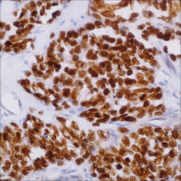
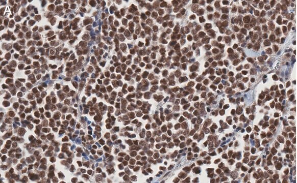
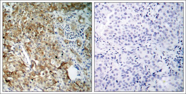
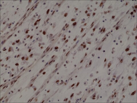
Anti-MYOD1 (Ab-200) antibody produced in rabbit

affinity isolated antibody

Synonym(s):

Anti-MYF3 antibody produced in rabbit, Anti-MYOD antibody produced in rabbit, Anti-PUM antibody produced in rabbit, Anti-bHLHc1 antibody produced in rabbit, Anti-myogenic differentiation 1 antibody produced in rabbit

Sign Into View Organizational & Contract Pricing


About This Item

MDL number:
UNSPSC Code:
12352203
NACRES:
NA.41

biological source

rabbit

conjugate

unconjugated

antibody form

affinity isolated antibody

antibody product type

primary antibodies

clone

polyclonal

form

buffered aqueous solution

mol wt

~40 kDa

species reactivity

mouse, human, rat

concentration

1 mg/mL

technique(s)

western blot: 1:500-1:1000

isotype

IgG

immunogen sequence

(A-S-S-P-R)

NCBI accession no.

UniProt accession no.

shipped in

wet ice

storage temp.

−20°C

target post-translational modification

unmodified

Gene Information

human ... MYOD1(4654)

General description

MYOD1 (myogenic differentiation 1) gene codes for a nuclear protein, that is a member of the basic helix-loop-helix (bHLH) family of transcription factors and the myogenic factors subfamily. It is located on human chromosome 11p15.1.

Immunogen

Peptide sequence around aa. 198!202 (A-S-S-P-R), according to the protein MYOD1.

Biochem/physiol Actions

Lack of MyoD (myogenic differentiation) results in a perinatally lethal fetal akinesia. It brings histone-modifying enzymes, which help to induce muscle differentiation.

Features and Benefits

Evaluate our antibodies with complete peace of mind. If the antibody does not perform in your application, we will issue a full credit or replacement antibody. Learn more.

Target description

MyoD encodes a nuclear protein that belongs to the basic helix-loop-helix family of transcription factors and the myogenic factors subfamily. It regulates muscle cell differentiation by inducing cell cycle arrest, a prerequisite for myogenic initiation. The protein is also involved in muscle regeneration. It activates its own transcription which may stabilize commitment to myogenesis.

Physical form

Solution in phosphate-buffered saline containing 0.02% sodium azide and 50% glycerol

Disclaimer

Unless otherwise stated in our catalog or other company documentation accompanying the product(s), our products are intended for research use only and are not to be used for any other purpose, which includes but is not limited to, unauthorized commercial uses, in vitro diagnostic uses, ex vivo or in vivo therapeutic uses or any type of consumption or application to humans or animals.

Not finding the right product?  

Try our Product Selector Tool.

Storage Class Code

10 - Combustible liquids

WGK

WGK 1

Flash Point(F)

Not applicable

Flash Point(C)

Not applicable


Certificates of Analysis (COA)

Search for Certificates of Analysis (COA) by entering the products Lot/Batch Number. Lot and Batch Numbers can be found on a product’s label following the words ‘Lot’ or ‘Batch’.

Already Own This Product?

Find documentation for the products that you have recently purchased in the Document Library.

Visit the Document Library

A recurrent neomorphic mutation in MYOD1 defines a clinically aggressive subset of embryonal rhabdomyosarcoma associated with PI3K-AKT pathway mutations
Kohsaka S, et al.
Nature Genetics, 46(6), 595-600 (2014)
Deficiency of the myogenic factor MyoD causes a perinatally lethal fetal akinesia
Watson CM, et al.
Journal of medical Genetics, 53(4), 264-269 (2016)
Manuela Dos Santos et al.
Advances in rheumatology (London, England), 62(1), 27-27 (2022-07-23)
Clinical evidence of skeletal muscle involvement is not uncommon in systemic lupus erythematosus (SLE). Because of the poor understanding of signaling pathways involved in SLE muscle wasting, the  aim of this study was to evaluate the effects of vitamin D supplementation
Narasimhan P Agaram et al.
Genes, chromosomes & cancer, 53(9), 779-787 (2014-05-16)
Sclerosing and spindle cell rhabdomyosarcoma (RMS) are rare types of RMS recently reclassified as a stand-alone pathologic entity, separate from embryonal RMS (ERMS). Although sclerosing and spindle cell RMS share clinical and morphologic features, a pathogenetic link based on shared
J Spring
FEBS letters, 400(1), 2-8 (1997-01-02)
For the growing fraction of human genes with identified functions there are often homologues known from invertebrates such as Drosophila. A survey of well established gene families from aldolases to zinc finger transcription factors reveals that usually a single invertebrate

Our team of scientists has experience in all areas of research including Life Science, Material Science, Chemical Synthesis, Chromatography, Analytical and many others.

Contact Technical Service