Skip to Content
Merck
All Photos(3)

Key Documents

E8655

Sigma-Aldrich

Anti-E6AP antibody, Mouse monoclonal

clone E6AP-330, purified from hybridoma cell culture

Synonym(s):

Anti-E2F-6

Sign Into View Organizational & Contract Pricing


About This Item

MDL number:
UNSPSC Code:
12352203
NACRES:
NA.41

biological source

mouse

Quality Level

conjugate

unconjugated

antibody form

purified from hybridoma cell culture

antibody product type

primary antibodies

clone

E6AP-330, monoclonal

form

buffered aqueous solution

mol wt

antigen ~100 kDa

species reactivity

human, mouse, rat, monkey

technique(s)

immunocytochemistry: suitable
immunoprecipitation (IP): suitable
indirect ELISA: suitable
microarray: suitable
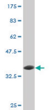
western blot: 1-2 μg/mL using total cell extract from 293T cells

isotype

IgG1

UniProt accession no.

shipped in

dry ice

storage temp.

−20°C

target post-translational modification

unmodified

Gene Information

human ... E2F6(1876)
mouse ... E2f6(50496)
rat ... E2f6(313978)

General description

E6AP is an E3 ubiquitin ligase that is expressed by the UB3A gene. Inhibiton or alterations of the UB3A gene may cause a neurological disorder called the Angelman Syndrome. E6AP interacts with E1 and E2 enzymes to mediate ubiquitination of proteins marked for degradation. E6AP also binds with the E6 viral protein present in HPV-infected cells.
Monoclonal Anti-E6AP antibody is a useful tool for the study of E6AP and its function in protein degradation. This antibody is specific for E6AP protein in rat, mouse, human and monkey.

Immunogen

human full-length recombinant E6AP.

Application

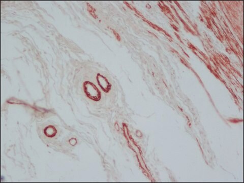
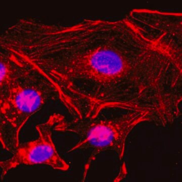
Monoclonal Anti-E6AP antibody is suitable for use in western blot (1-2 μg/mL using total cell extract from 293T cells), immunocytochemistry, immunoblotting, immunoprecipitation, indirect ELISA and microarray.

Physical form

Solution in 0.01 M phosphate buffered saline, pH 7.4, containing 15 mM sodium azide.

Disclaimer

Unless otherwise stated in our catalog or other company documentation accompanying the product(s), our products are intended for research use only and are not to be used for any other purpose, which includes but is not limited to, unauthorized commercial uses, in vitro diagnostic uses, ex vivo or in vivo therapeutic uses or any type of consumption or application to humans or animals.

Not finding the right product?  

Try our Product Selector Tool.

Storage Class Code

10 - Combustible liquids

WGK

nwg

Flash Point(F)

Not applicable

Flash Point(C)

Not applicable


Certificates of Analysis (COA)

Search for Certificates of Analysis (COA) by entering the products Lot/Batch Number. Lot and Batch Numbers can be found on a product’s label following the words ‘Lot’ or ‘Batch’.

Already Own This Product?

Find documentation for the products that you have recently purchased in the Document Library.

Visit the Document Library

Tiantian Wang et al.
Frontiers in cellular neuroscience, 11, 418-418 (2018-01-23)
The neurodevelopmental disorder Angelman syndrome (AS) is characterized by intellectual disability, motor dysfunction, distinct behavioral aspects, and epilepsy. AS is caused by a loss of the maternally expressed UBE3A gene, and many of the symptoms are recapitulated in a Ube3a
The role of TP53 in Cervical carcinogenesis.
Tommasino, M., et al.
Human Mutation, 21, 307-312 (2003)
Yi Sun
Cancer biology & therapy, 2(6), 623-629 (2003-12-23)
E3 ubiquitin ligases are a large family of proteins that can be classified into three major structurally distinct types: N-end rule E3s, E3s containing the HECT (Homology to E6AP C-Terminus) domain, and E3s with the RING (Really Interesting New Gene)
Stuti Chhabra et al.
Biochimica et biophysica acta, 1864(10), 1545-1553 (2017-06-06)
Granulocyte colony-stimulating factor receptor (G-CSFR) plays a crucial role in regulating myeloid cell survival, proliferation, and neutrophilic granulocyte precursor cells maturation. Previously, we demonstrated that Fbw7α negatively regulates G-CSFR and its downstream signaling through ubiquitin-proteasome mediated degradation. However, whether additional
Sheng Miao et al.
The Journal of neuroscience : the official journal of the Society for Neuroscience, 33(1), 327-333 (2013-01-04)
Pyramidal neurons have a highly polarized dendritic morphology, characterized by one long apical dendrite and multiple short basal dendrites. They function as the primary excitatory cells of the mammalian prefrontal cortex and the corticospinal tract. However, the molecular mechanisms underlying

Our team of scientists has experience in all areas of research including Life Science, Material Science, Chemical Synthesis, Chromatography, Analytical and many others.

Contact Technical Service