Skip to Content
Merck
All Photos(1)

Documents

156M-8

Sigma-Aldrich

CD56 (123C3.D5) Mouse Monoclonal Antibody

clone 123C3.D5, unconjugated, Cell Marque

Sign Into View Organizational & Contract Pricing


About This Item

UNSPSC Code:
12352203
NACRES:
NA.41

biological source

mouse

Quality Level

100
500

conjugate

unconjugated

antibody form

culture supernatant

antibody product type

primary antibodies

clone

123C3.D5, monoclonal

description

For In Vitro Diagnostic Use in Select Regions (See Chart)

form

buffered aqueous solution

species reactivity

human

packaging

vial of 0.1 mL concentrate (156M-84)
vial of 0.5 mL concentrate (156M-85)
bottle of 1.0 mL predilute (156M-87)
vial of 1.0 mL concentrate (156M-86)
bottle of 7.0 mL predilute (156M-88)

manufacturer/tradename

Cell Marque

technique(s)

immunohistochemistry (formalin-fixed, paraffin-embedded sections): 1:100-1:500

isotype

IgG1κ

control

neuroblastoma

shipped in

wet ice

storage temp.

2-8°C

visualization

membranous

Gene Information

human ... NCAM1(4684)

General description

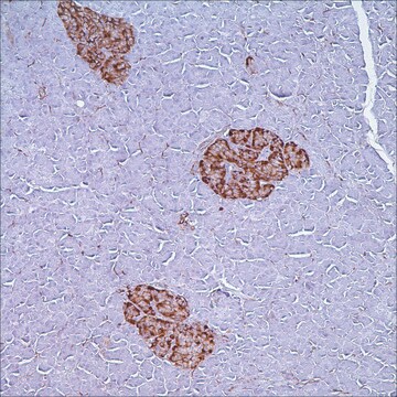
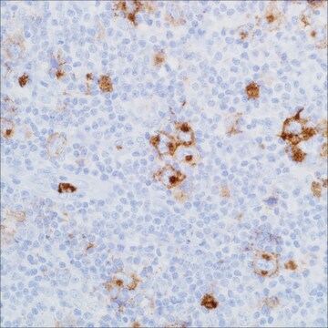
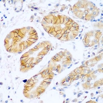
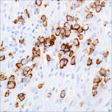
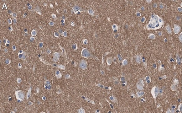
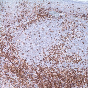
Anti-CD56 recognizes two proteins of the neural cell adhesion molecule, the basic molecule expressed on most neuroectodermally derived cell lines, tissues and neoplasms (e.g. retinoblastoma, medulloblastomas, astrocytomas, neuroblastomas, and small cell carcinomas). It is also expressed on some mesodermally derived tumors (rhabdomyosarcoma).

Linkage

CD56 Positive Control Slides, Product No. 156S, are available for immunohistochemistry (formalin-fixed, paraffin-embedded sections).

Physical form

Solution in Tris Buffer, pH 7.3-7.7, with 1% BSA and <0.1% Sodium Azide

Preparation Note

Download the IFU specific to your product lot and formatNote: This requires a keycode which can be found on your packaging or product label.

Other Notes

For Technical Service please contact: 800-665-7284 or email: service@cellmarque.com

Legal Information

Cell Marque is a trademark of Merck KGaA, Darmstadt, Germany

Not finding the right product?  

Try our Product Selector Tool.


Certificates of Analysis (COA)

Search for Certificates of Analysis (COA) by entering the products Lot/Batch Number. Lot and Batch Numbers can be found on a product’s label following the words ‘Lot’ or ‘Batch’.

Already Own This Product?

Find documentation for the products that you have recently purchased in the Document Library.

Visit the Document Library

R Gerardy-Schahn et al.
International journal of cancer. Supplement = Journal international du cancer. Supplement, 8, 38-42 (1994-01-01)
Monoclonal antibodies (MAbs) ranked together as small-cell-lung-cancer (SCLC) Cluster I MAbs are directed against the neural cell adhesion molecule NCAM (CD 56) and have been shown to be useful reagents in SCLC diagnosis and therapy. We analyzed the epitopes recognized
C E Moolenaar et al.
Cancer research, 50(4), 1102-1106 (1990-02-15)
Monoclonal antibodies (MAbs) 123C3 and 123A8 generated against a membrane preparation of a small cell lung carcinoma (SCLC) specimen recognize not only SCLC and bronchial carcinoids but also a significant portion of non-small cell lung carcinomas (non-SCLC) of various histological
R E Kibbelaar et al.
European journal of cancer (Oxford, England : 1990), 27(4), 431-435 (1991-01-01)
We investigated the expression of the neural cell adhesion molecule (NCAM) in a series of surgically resected lung carcinomas of various histological subtypes by means of a panel of monoclonal antibodies recognising different N-CAM epitopes. In a subgroup of 56
Scott A Ely et al.
The American journal of pathology, 160(4), 1293-1299 (2002-04-12)
Unlike monoclonal gammopathy of undetermined significance (MGUS) or non-Hodgkin's lymphomas (NHLs) with plasmacytoid differentiation, multiple myeloma (MM) is commonly associated with lytic bone lesions. Although the mechanisms of increased osteoclast activity are partially understood, comparatively little is known about the
Masahiko Sumi et al.
Leukemia & lymphoma, 44(1), 201-204 (2003-04-15)
We present a case of duodenal non-Hodgkin lymphoma in a 71-year-old woman. Immunohistochemistry characterized the lymphoma cells as CD2(+); surface CD3(-) but cytoplasmic CD3(+); CD7(+); and CD56(+) without a rearrangement of the T-cell receptor gene. Cells had a high N/C

Our team of scientists has experience in all areas of research including Life Science, Material Science, Chemical Synthesis, Chromatography, Analytical and many others.

Contact Technical Service