Skip to Content
Merck
All Photos(3)

Key Documents

MABN2280

Sigma-Aldrich

Anti-FMR1polyG Antibody, clone 8FM-2F7

ascites fluid, clone 8FM-2F7, from mouse

Synonym(s):

Synaptic functional regulator FMR1, Fragile X mental retardation protein 1, FMRP, Protein FMR-1

Sign Into View Organizational & Contract Pricing


About This Item

UNSPSC Code:
12352203
eCl@ss:
32160702

biological source

mouse

antibody form

ascites fluid

antibody product type

primary antibodies

clone

8FM-2F7, monoclonal

species reactivity

human

packaging

antibody small pack of 25 μL

technique(s)

immunofluorescence: suitable
immunohistochemistry: suitable (paraffin)
western blot: suitable

isotype

IgG1κ

NCBI accession no.

UniProt accession no.

target post-translational modification

unmodified

Gene Information

human ... FMR1(2332)

General description

Synaptic functional regulator FMR1 (UniProt: Q06787; also known as Fragile X mental retardation protein 1, FMRP, Protein FMR-1) is encoded by the FMR1 gene (Gene ID: 2332) in human. FMR-1 is a multifunctional polyribosome-associated RNA-binding protein that plays a central role in neuronal development and synaptic plasticity through the regulation of alternative mRNA splicing, mRNA stability, mRNA dendritic transport and postsynaptic local protein synthesis of a subset of mRNAs. It plays a role in mRNA nuclear export and together with export factor NXF2, it is involved in the regulation of the NXF1 mRNA stability in neurons. It is also reported to stabilize the scaffolding postsynaptic density protein DLG4/PSD-95 and the myelin basic protein mRNAs in hippocampal neurons and glial cells, respectively and this stabilization is further increased in response to metabotropic glutamate receptor stimulation. A CGG repeat expansion (55-200) in the 5 UTR of FMR1 gene is reported to cause Fragile X-associated Tremor Ataxia Syndrome (FXTAS). The expanded CGG repeat is associated with elevated FMR1 mRNA expression that results and neurodegeneration. The CGG repeat expansion triggers repeat-associated non-ATG-initiated translation (RAN or RANT) within the 5 UTR of FMR1 mRNA, resulting in the production of fusion proteins (FMR1polyG) that contain N-terminal polyglycine fused to either C-terminal FMR1 in-frame sequence or one of two FMR1 frame-shift sequences. The FMR1polyG-positive aggregates closely resemble neuronal intranuclear inclusions seen in polyglutamine diseases and other protein-mediated neurodegenerative disorders. (Ref.: Buijsen, RAM., et al. (2014). Acta Neuropathol. Commun. 2:162; Todd, PK et al. (2013). Neuron. 78(3); 440-455).

Specificity

Clone 8FM-2F7 specifically detects FMR1polyG in human cells.

Immunogen

Epitope: N-terminus
Ovalbumin-conjugated linear peptide with EAPLPGGVRQRGG sequence. Note: This protein is translated within the 5′UTR of the FMR1 gene and in a different frame resulting in a protein with different sequence and containing polyGlycine.

Application

Anti-FMR1polyG, clone 8FM-2F7, Cat. No. MABN2280, is a highly specific mouse monoclonal antibody that targets Synaptic functional regulator FMR1 (FMR1polyG) and has been tested for use Immunofluorescence, Immunohistochemistry (Paraffin), and Western Blotting.
Research Category
Neuroscience
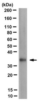
Western Blotting Analysis: A 1:500 dilution from a representative lot detected FMR1polyG in lysate from HEK293 cells overexpressing FMR1polyG.

Immunohistochemistry Analysis: A representative lot detected FMR1polyG in Immunohistochemistry applications (Buijsen, R.A., et. al. (2014). Acta Neuropathol Commun. 2:162; Sellier, C., et. al. (2017). Neuron. 93(2):331-347).

Western Blotting Analysis: A representative lot detected FMR1polyG in Western Blotting applications (Sellier, C., et. al. (2017). Neuron. 93(2):331-347).

Immunohistochemistry Analysis: A 1:50 dilution from a representative lot detected FMR1polyG in human cerebral cortex and human testis tissues.

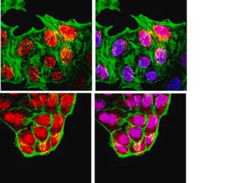
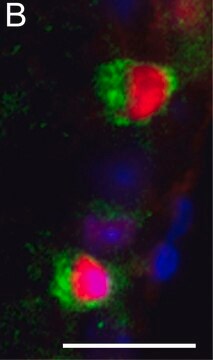
Immunofluorescence Analysis: A representative lot detected FMR1polyG in Immunofluorescence applications (Buijsen, R.A., et. al. (2014). Acta Neuropathol Commun. 2:162; Sellier, C., et. al. (2017). Neuron. 93(2):331-347).

Quality

Evaluated by Western Blotting in HEK293 cell lysates expressing FMR1polyG.

Western Blotting Analysis: A 1:500 dilution of this antibody detected FMR1polyG in HEK293 cell lysates expressing FMR1polyG.

Target description

~35 kDa observed. Uncharacterized bands may be observed in some lysate(s).

Physical form

Mouse monoclonal antibody in ascites without preservatives.
Unpurified

Storage and Stability

Stable for 1 year at -20°C from date of receipt. Handling Recommendations: Upon receipt and prior to removing the cap, centrifuge the vial and gently mix the solution. Aliquot into microcentrifuge tubes and store at -20°C. Avoid repeated freeze/thaw cycles, which may damage IgG and affect product performance.

Other Notes

Concentration: Please refer to lot specific datasheet.

Disclaimer

Unless otherwise stated in our catalog or other company documentation accompanying the product(s), our products are intended for research use only and are not to be used for any other purpose, which includes but is not limited to, unauthorized commercial uses, in vitro diagnostic uses, ex vivo or in vivo therapeutic uses or any type of consumption or application to humans or animals.

Not finding the right product?  

Try our Product Selector Tool.


Certificates of Analysis (COA)

Search for Certificates of Analysis (COA) by entering the products Lot/Batch Number. Lot and Batch Numbers can be found on a product’s label following the words ‘Lot’ or ‘Batch’.

Already Own This Product?

Find documentation for the products that you have recently purchased in the Document Library.

Visit the Document Library

Our team of scientists has experience in all areas of research including Life Science, Material Science, Chemical Synthesis, Chromatography, Analytical and many others.

Contact Technical Service