17-10230
LentiBrite GFP-LC3-II Enrichment Kit (Flow Cytometry)
Sign Into View Organizational & Contract Pricing
All Photos(1)
About This Item
UNSPSC Code:
12161503
eCl@ss:
32161000
NACRES:
NA.84
Recommended Products
General description
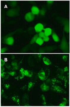
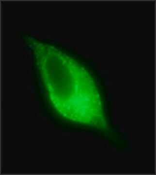
Autophagy, a degradative pathway that provides recycled nutrients to cells under stress, plays both protective and deleterious roles in many diseases, including cancer, neurodegeneration, and infections. Members of the LC3 family play a key role in the maturation of the autophagosome, the central organelle of autophagy. LC3 precursors, diffusely distributed in the cytosol, are proteolytically processed to form LC3-I. Upon initiation of autophagy, the C-terminal glycine is modified by addition of a phosphatidylethanolamine (PE) to form LC3-II, which translocates rapidly to nascent autophagosomes in a punctate distribution. DNA constructs encoding fluorescent proteins fused to LC3 are widely employed for introduction into cells for monitoring autophagosome formation by fluorescence microscopy. <br />EMD Millipore′s LentiBrite GFP-LC3 lentiviral particles provide bright fluorescence and precise localization to enable live cell analysis of autophagy in difficult-to-transfect cell types. The <br /><br />LentiBrite GFP-LC3-II Enrichment Kit (Flow Cytometry) combines our GFP-LC3 lentiviral particles with a selective permeabilization procedure for GFP-LC3 transduced cells that allows release of cytosolic LC3-I and retention of autophagosome-bound LC3-II. This procedure allows detection of autophagosome density by flow cytometry.
Application
EMD Millipore′s LentiBrite GFP-LC3 lentiviral particles provide bright fluorescence and precise localization to enable live cell analysis of autophagy in difficult-to-transfect cell types. The LentiBrite GFP-LC3-II Enrichment Kit (Flow Cytometry) combines our GFP-LC3 lentiviral particles with a selective permeabilization procedure for GFP-LC3 transduced cells that allows release of cytosolic LC3-I and retention of autophagosome-bound LC3-II. This procedure allows detection of autophagosome density by flow cytometry.
For optimal fluorescent analysis, it is recommended to analyze the target expression level within 24-48 hrs after transfection/infection for optimal live cell analysis, as fluorescent intensity may dim over time, especially in difficult-to-transfect cell lines. Infected cells may be frozen down after successful transfection/infection and thawed in culture to retain positive fluorescent expression beyond 24-48 hrs. Length and intensity of fluorescent expression varies between cell lines. Higher MOIs may be required for difficult-to-transfect cell lines.
For optimal fluorescent analysis, it is recommended to analyze the target expression level within 24-48 hrs after transfection/infection for optimal live cell analysis, as fluorescent intensity may dim over time, especially in difficult-to-transfect cell lines. Infected cells may be frozen down after successful transfection/infection and thawed in culture to retain positive fluorescent expression beyond 24-48 hrs. Length and intensity of fluorescent expression varies between cell lines. Higher MOIs may be required for difficult-to-transfect cell lines.
Components
1. TagGFP2-LC3 Lentivirus: One vial containing 25 µL of lentiviral particles at a minimum of 3 x 10E8 infectious units (IFU) per mL. For lot-specific titer information, please see datasheet. • Virus Description: 3rd generation HIV-based VSV-G pseudotyped lentiviral particles. Lentiviral particles were generated using the pPACKH1 Lentivector Packaging System and purified by PEG precipitation. • Promoter: EF-1 (Elongation Factor-1). • Expression Marker Description: TagGFP2 (Evrogen) fused at its C-terminus with human LC3A (Accession # NP_115903) truncated to leave G120 (lipidation site) at the C-terminus. (Store at -80°C) 2. Autophagy Reagent A: One vial (lyophilized) (Store at 2-8°C) 3. 10x Enrichment Buffer (Part No. CS210552): One vial containing 1 mL (Store at 2-8°C) 4. 5x Assay Buffer: (Part No. CS210529): One bottle containing 3 mL (Store at 2-8°C)
Storage and Stability
Store TagGFP2-LC3 Lentivirus at -80°C. After first thaw, place immediately on ice and freeze in working aliquots at -80°C. Frozen aliquots may be stored for at least 2 months. Further freeze/thaws may result in decreased virus titer and transduction efficiency. Store Autophagy Reagent A, 10x Enrichment Buffer, and 5x Assay Buffer at 2-8°C. All components are stable for at least 4 months from date of receipt when stored as recommended above.
Disclaimer
This product contains genetically modified organisms (GMO). Within the EU GMOs are regulated by Directives 2001/18/EC and 2009/41/EC of the European Parliament and of the Council and their national implementation in the member States respectively. This legislation obliges {HCompany} to request certain information about you and the establishment where the GMOs are being handled. Click here for Enduser Declaration (EUD) Form.Unless otherwise stated in our catalog or other company documentation accompanying the product(s), our products are intended for research use only and are not to be used for any other purpose, which includes but is not limited to, unauthorized commercial uses, in vitro diagnostic uses, ex vivo or in vivo therapeutic uses or any type of consumption or application to humans or animals.
Signal Word
Warning
Hazard Statements
Precautionary Statements
Hazard Classifications
Acute Tox. 4 Oral - Aquatic Chronic 3
Storage Class Code
10 - Combustible liquids
WGK
WGK 3
Certificates of Analysis (COA)
Search for Certificates of Analysis (COA) by entering the products Lot/Batch Number. Lot and Batch Numbers can be found on a product’s label following the words ‘Lot’ or ‘Batch’.
Already Own This Product?
Find documentation for the products that you have recently purchased in the Document Library.
Our team of scientists has experience in all areas of research including Life Science, Material Science, Chemical Synthesis, Chromatography, Analytical and many others.
Contact Technical Service
