Skip to Content
Merck
All Photos(2)

Key Documents

SAB4500369

Sigma-Aldrich

Anti-Collagen IV antibody produced in rabbit

affinity isolated antibody

Synonym(s):

Arresten, COL4A1, Collagen α-1(IV) chain

Sign Into View Organizational & Contract Pricing


About This Item

UNSPSC Code:
12352203
NACRES:
NA.41

biological source

rabbit

Quality Level

conjugate

unconjugated

antibody form

affinity isolated antibody

antibody product type

primary antibodies

clone

polyclonal

form

buffered aqueous solution

mol wt

antigen 160 kDa

species reactivity

human, mouse

concentration

~1 mg/mL

technique(s)

ELISA: 1:20000
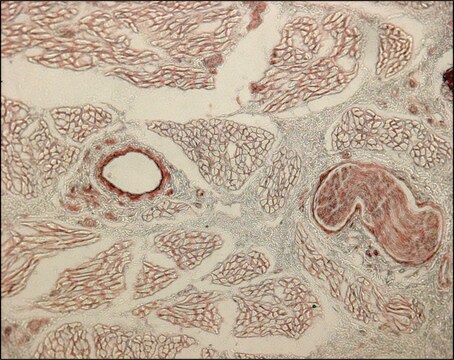
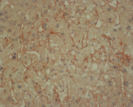
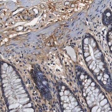
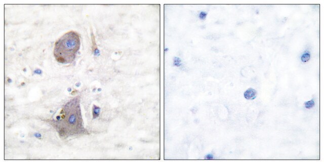
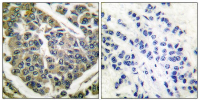
immunohistochemistry: 1:50-1:100
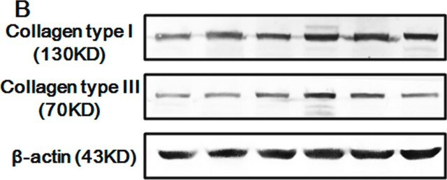
western blot: 1:500-1:1000

NCBI accession no.

UniProt accession no.

shipped in

wet ice

storage temp.

−20°C

target post-translational modification

unmodified

Gene Information

human ... COL4A1(1282)

General description

Anti-Collagen IV Antibody detects endogenous levels of total Collagen IV protein.
COL4A1 (collagen type IV alpha 1 chain) belongs to the collagen family. It has an N-terminal 7S domain, a C-terminal globular domain (NC1), and the central triple helical part with short interruptions of the Gly-X-Y repeats. It is an important constituent of type IV collagen in the basement membrane. The COL4A1 gene is located on human chromosome 13q34.

Immunogen

The antiserum was produced against synthesized peptide derived from human Collagen IV.

Immunogen Range: 11-60

Application

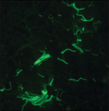
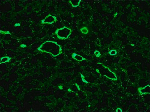
Anti-Collagen IV, N-Terminal antibody has been used in histology analysis, immunofluorescence microscopy and immunostaining.

Biochem/physiol Actions

COL4A1 (collagen type IV α 1 chain) related disorder is associated with pulmonary complication. This gene is a major constituent required in basal membrane stability. Mutations in COL4A1 result in familial porencephaly, infantile hemiplegia, cerebral small vessel disease (CSVD) and hemorrhagic stroke.

Features and Benefits

Evaluate our antibodies with complete peace of mind. If the antibody does not perform in your application, we will issue a full credit or replacement antibody. Learn more.

Physical form

Rabbit IgG in phosphate buffered saline (without Mg2+ and Ca2+), pH 7.4, 150mM NaCl, 0.02% sodium azide and 50% glycerol.

Disclaimer

Unless otherwise stated in our catalog or other company documentation accompanying the product(s), our products are intended for research use only and are not to be used for any other purpose, which includes but is not limited to, unauthorized commercial uses, in vitro diagnostic uses, ex vivo or in vivo therapeutic uses or any type of consumption or application to humans or animals.

Not finding the right product?  

Try our Product Selector Tool.

Storage Class Code

10 - Combustible liquids

WGK

nwg

Flash Point(F)

Not applicable

Flash Point(C)

Not applicable


Certificates of Analysis (COA)

Search for Certificates of Analysis (COA) by entering the products Lot/Batch Number. Lot and Batch Numbers can be found on a product’s label following the words ‘Lot’ or ‘Batch’.

Already Own This Product?

Find documentation for the products that you have recently purchased in the Document Library.

Visit the Document Library

Visualization of basement membranes in normal breast and breast cancer tissues using multiphoton microscopy.
Wu X, et al.
Oncology Letters, 11(6), 3785-3789 (2016)
Dongbo Zhao et al.
Endocrine journal, 68(7), 751-762 (2021-04-02)
Rosiglitazone (RSG) is widely used to reduce the amount of sugar in the blood of patients with diabetes mellitus. Diabetic nephropathy is the most common microvascular complication of diabetes. The role of RSG in diabetic nephropathy is not fully understood.
Ramesh B Kasetti et al.
The Journal of biological chemistry, 293(25), 9854-9868 (2018-05-11)
Elevation of intraocular pressure (IOP) is a serious adverse effect of glucocorticoid (GC) therapy. Increased extracellular matrix (ECM) accumulation and endoplasmic reticulum (ER) stress in the trabecular meshwork (TM) is associated with GC-induced IOP elevation. However, the molecular mechanisms by
Effects of conventional immunosuppressive therapy on functional and pathological features of CNS lupus in NZB/W mice.
Alexander J, et al.
F1000Research, 4 (2018)
Xiufeng Wu et al.
Oncology letters, 11(6), 3785-3789 (2016-06-18)
Since basement membranes represent a critical barrier during breast cancer progression, timely imaging of these signposts is essential for early diagnosis of breast cancer. A label-free method using multiphoton microscopy (MPM) based on two-photon excited fluorescence signals and second harmonic

Our team of scientists has experience in all areas of research including Life Science, Material Science, Chemical Synthesis, Chromatography, Analytical and many others.

Contact Technical Service