S8902
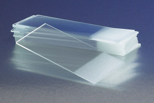
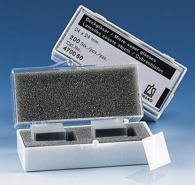
Slides, microscope
plain, size 25 mm × 75 mm
Synonym(s):
glass slide
About This Item
Recommended Products
material
glass
packaging
case of 20
box of 72 slides
size
25 mm × 75 mm
binding type
non-treated surface
Looking for similar products? Visit Product Comparison Guide
General description
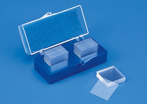
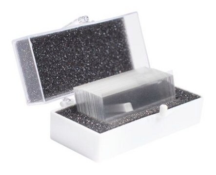
Our microscope slides are made of high-quality white glass with low iron content, no green tint with fine texture and transparency; these slides are also corrosion-resistant. The edges are ground smooth to help eliminate sharp cutting surfaces and safe handling. They are packed in a special thermoform plastic box to minimize exposure to moisture and dust (each slide is precleaned).
Conform to Federal specification NNN-S-450.
Certificates of Analysis (COA)
Search for Certificates of Analysis (COA) by entering the products Lot/Batch Number. Lot and Batch Numbers can be found on a product’s label following the words ‘Lot’ or ‘Batch’.
Already Own This Product?
Find documentation for the products that you have recently purchased in the Document Library.
Protocols
Cell staining can be divided into four steps: cell preparation, fixation, application of antibody, and evaluation.
Related Content
Three-dimensional (3D) printing of biological tissue is rapidly becoming an integral part of tissue engineering.
Our team of scientists has experience in all areas of research including Life Science, Material Science, Chemical Synthesis, Chromatography, Analytical and many others.
Contact Technical Service