Skip to Content
Merck
All Photos(1)

Key Documents

B6529

Sigma-Aldrich

Brilliant Blue R Staining Solution

ethanol solution

Synonym(s):

Brilliant Blue R, Acid Blue 83, Brilliant indocyanin 6B, Coomassie Brilliant Blue R

Sign Into View Organizational & Contract Pricing


About This Item

Empirical Formula (Hill Notation):
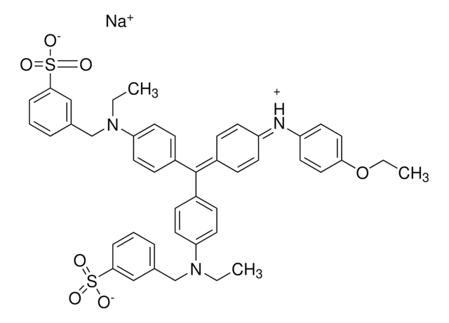
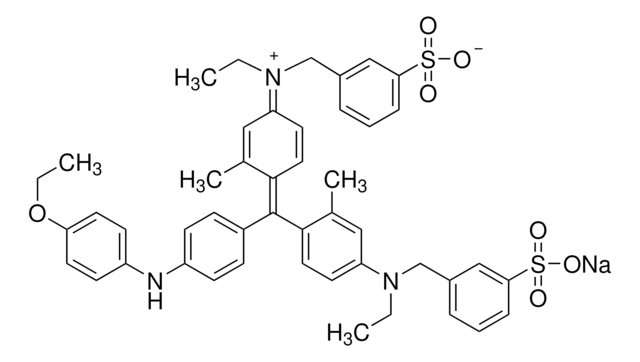
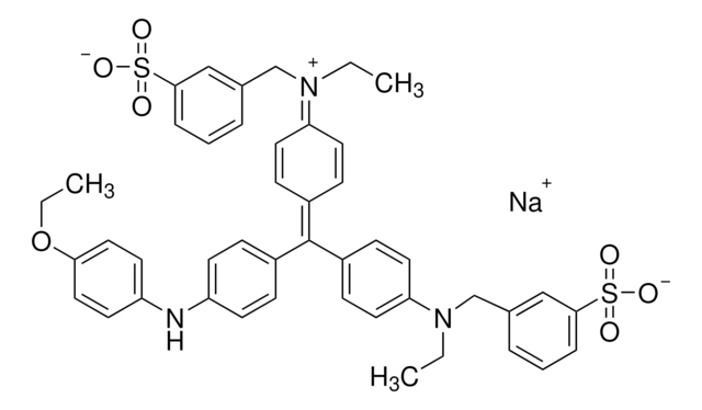
C45H44N3NaO7S2
CAS Number:
Molecular Weight:
825.97
Colour Index Number:
42660
Beilstein:
5718025
MDL number:
UNSPSC Code:
12171500
PubChem Substance ID:
NACRES:
NA.47

product name

Brilliant Blue R Staining Solution, suitable for (for immunoelectrophoresis protein staining)

form

liquid

Quality Level

technique(s)

microbe id | staining: suitable

color

dark blue

suitability

suitable for (for immunoelectrophoresis protein staining)

application(s)

diagnostic assay manufacturing
hematology
histology

storage temp.

room temp

SMILES string

[Na+].CCOc1ccc(Nc2ccc(cc2)C(\c3ccc(cc3)N(CC)Cc4cccc(c4)S([O-])(=O)=O)=C5\C=C/C(C=C5)=[N+](\CC)Cc6cccc(c6)S([O-])(=O)=O)cc1

InChI

1S/C45H45N3O7S2.Na/c1-4-47(31-33-9-7-11-43(29-33)56(49,50)51)40-23-15-36(16-24-40)45(35-13-19-38(20-14-35)46-39-21-27-42(28-22-39)55-6-3)37-17-25-41(26-18-37)48(5-2)32-34-10-8-12-44(30-34)57(52,53)54;/h7-30H,4-6,31-32H2,1-3H3,(H2,49,50,51,52,53,54);/q;+1/p-1

InChI key

NKLPQNGYXWVELD-UHFFFAOYSA-M

Looking for similar products? Visit Product Comparison Guide

Application

Brilliant Blue R staining solution is especially designed for use in staining protein in agarose gels following immunoelectrophoresis and Ouchterlony gels. The stain contains ethanol and acetic acid so gels do not require fixing prior to staining. The immunoelectrophoresis or Ouchterlony gel is first washed with water and saline to remove non-precipitated proteins and then dried. The gel is then immersed in staining solution for 30 min and destained with 10% acetic acid.
For detection of protein bands following electrophoresis.

Components

0.5% (w/v) Brilliant Blue R, 45% (v/v) ethanol and 10% (v/v) acetic acid.

Analysis Note

Tested for suitability on agarose gels following immunoelectrophoresis.

Legal Information

Pictograms

FlameExclamation mark

Signal Word

Warning

Hazard Statements

Hazard Classifications

Eye Irrit. 2 - Flam. Liq. 3 - Skin Irrit. 2

Storage Class Code

3 - Flammable liquids

WGK

WGK 2

Flash Point(F)

81.0 °F - closed cup

Flash Point(C)

27.2 °C - closed cup

Personal Protective Equipment

dust mask type N95 (US), Eyeshields, Gloves

Choose from one of the most recent versions:

Certificates of Analysis (COA)

Lot/Batch Number

Don't see the Right Version?

If you require a particular version, you can look up a specific certificate by the Lot or Batch number.

Already Own This Product?

Find documentation for the products that you have recently purchased in the Document Library.

Visit the Document Library

Wei Zhang et al.
International journal of oncology, 54(5), 1719-1733 (2019-03-14)
Ovarian cancer remains the most lethal type of cancer among all gynecological malignancies. The majority of patients are diagnosed with ovarian cancer at the late stages of the disease. Therefore, there exists an imperative need for the development of early
K Kubo
Analytical biochemistry, 213(2), 200-205 (1993-09-01)
In sodium dodecyl sulfate-polyacrylamide gel electrophoresis (SDS-PAGE) according to the method of Weber and Osborn, protein bands are often distorted by tailing at both ends, and the phenomenon is often called "edge tailing." This was eliminated by adding glycerol at
Zhang Xin-Guo Chen Jian-Hua et al.
Journal of chromatographic science, 50(9), 820-825 (2012-06-22)
A human serum albumin and Thymosin α1 (HSA-Tα1) fusion protein was designed and over-expressed in Pichia pastoris. To purify the fusion protein, a new native preparative electrophoresis system that involved a modified device with a sample receiving chamber, and an
Reiner Westermeier
Proteomics, 6 Suppl 2, 61-64 (2006-10-13)
In spite of the high sensitivity of silver staining and the wide dynamic range of various fluorescent detection methods, Coomassie Brilliant Blue staining is still the most widely used protein detection technique for proteins separated by polyacrylamide gel electrophoresis. There
Chun Yi Liau et al.
Journal of bioscience and bioengineering, 106(1), 111-113 (2008-08-12)
A modified Coomassie Brilliant Blue G 250 staining method for detecting chitinolytic enzymes in chitin-containing polyacrylamide gel electrophoresis (PAGE) is presented. The staining formed achromatic zones at the locations of the migrated enzyme. Using Streptomyces griseus chitinase, we have demonstrated

Articles

MISSION® Target ID Library for Human miRNA Target Identification and Discovery;

To meet the great diversity of protein analysis needs, Sigma offers a wide selection of protein visualization (staining) reagents. EZBlue™ and ProteoSilver™, designed specifically for proteomics, also perform impressively in traditional PAGE formats.

Our team of scientists has experience in all areas of research including Life Science, Material Science, Chemical Synthesis, Chromatography, Analytical and many others.

Contact Technical Service