CELLDETH-RO
Roche
Cell Death Detection ELISAPLUS
sufficient for 96 multiwell tests (11774425001), sufficient for 10 x 96 multiwell tests (11920685001)
Synonym(s):
ELISA
About This Item
Recommended Products
usage
sufficient for 10 x 96 multiwell tests (11920685001)
sufficient for 96 multiwell tests (11774425001)
Quality Level
manufacturer/tradename
Roche
technique(s)
ELISA: suitable
detection method
photometric
storage temp.
2-8°C
General description
Specificity
Application
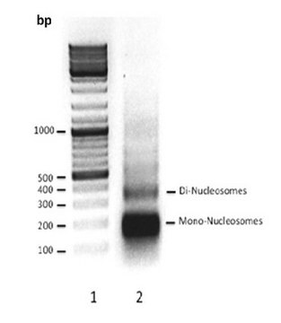
The Cell Death Detection ELISAPLUS photometric enzyme immunoassay is used for the quantitative in vitro determination of cytoplasmic histone-associated DNA fragments (mono- and oligonucleosomes) after induced cell death.
The kit contains a stop solution which allows users to terminate the substrate reaction and run the assay under defined conditions, making it suitable for use in high-throughput applications.
Features and Benefits
- One-step ELISA
- High sensitivity: (5 x 10² cells/ml)
- Fast performance: (3 - 4 hours)
- Positive control included
- No prelabeling of cells necessary
- Nonradioactive assay system
- No species restriction
- Easy handling
- Low background
- Suppressed human anti-mouse factor
- Function tested
Packaging
- 11774425001 (1 kit containing 10 components)
- 11920685001 (1 kit containing 10 components)
Specifications
Negative control: Depending on cell culture conditions, each exponentially growing permanent cell culture contains a certain amount of dead cells (typically approximately 3 - 8%). In the immunoassay, these inherent dead cells in the untreated sample (without a cell-death-inducing agent) will cause a certain absorbance value (negative control).
Positive control: A DNA-histone complex serves as a positive control.
Sample material: Cytoplasmic fractions (lysates) of cell lines, cells ex vivo, cell culture supernatants, and serum or plasma
Unit Definition
Preparation Note
Alongside the ready-to-use solutions supplied with this kit, you will need to prepare the following working solutions:
Note: Use only double-distilled water for reconstitution of lyophilizates.
Anti-Histone Biotin
Reconstitute the lyophilizate in 450 μl double-distilled water for 10 minutes and mix thoroughly.
Use: Component of the Immunoreagent.
Anti-DNA Peroxidase
Reconstitute the lyophilizate in 450 μl double-distilled water for 10 minutes and mix thoroughly.
Use: Component of the Immunoreagent.
Positive Control
Reconstitute the lyophilizate in 450 μl double-distilled water for 10 minutes and mix thoroughly.
Use: ELISA Step 1.
ABTS Tablets
Dependent on the number of samples tested, dissolve 1, 2, or 3 tablets from bottle 7 in 5, 10, or 15 ml Substrate Buffer.
Use: ELISA Step 5.
ABTS Stop Solution
If turbidity or a precipitate is visible, warm to 37 °C with shaking until the solution is clear.
Use: ELISA Step 6.
Storage conditions (working solution): Anti-Histone Biotin: 2 to 8 °C for 2 months
ABTS Tablets: 1 month stored protected from light. Allow to come to 15 to 25 °C before use.
ABTS Stop Solution: 2 to 8 °C until the expiration date printed on the label.
Other Notes
Kit Components Only
- Anti-histone-biotin antibody (clone H11-4)
- Anti-DNA-POD antibody (clone MCA-33)
- Positive Control
- Incubation Buffer
- Lysis Buffer
- Substrate Buffer
- ABTS Substrate Tablet
- ABTS Stop Solution
- Microplate (streptavidin-coated)
- Adhesive Cover Foils
Signal Word
WarningDanger
Hazard Statements
Hazard Classifications
Aquatic Chronic 2 - Aquatic Chronic 3 - Eye Dam. 1 - Eye Irrit. 2 - Skin Sens. 1
Storage Class Code
8B - Non-combustible, corrosive hazardous materials
WGK
WGK 3
Flash Point(C)
does not flash
Certificates of Analysis (COA)
Search for Certificates of Analysis (COA) by entering the products Lot/Batch Number. Lot and Batch Numbers can be found on a product’s label following the words ‘Lot’ or ‘Batch’.
Already Own This Product?
Find documentation for the products that you have recently purchased in the Document Library.
Customers Also Viewed
Articles
Cellular apoptosis assays to detect programmed cell death using Annexin V, Caspase and TUNEL DNA fragmentation assays.
Our team of scientists has experience in all areas of research including Life Science, Material Science, Chemical Synthesis, Chromatography, Analytical and many others.
Contact Technical Service

