Skip to Content
Merck
All Photos(2)

Documents

MABT118

Sigma-Aldrich

Anti-Desmoglein-1 Antibody, clone 32-2B

clone 32-2B, from mouse

Synonym(s):

Desmoglein-1, Desmosomal glycoprotein 1, DG1, DGI, Pemphigus foliaceus antigen

Sign Into View Organizational & Contract Pricing


About This Item

UNSPSC Code:
12352203
eCl@ss:
32160702
NACRES:
NA.41

biological source

mouse

Quality Level

antibody form

purified immunoglobulin

antibody product type

primary antibodies

clone

32-2B, monoclonal

species reactivity

human

species reactivity (predicted by homology)

bovine (immunogen homology)

technique(s)

immunocytochemistry: suitable
immunofluorescence: suitable
western blot: suitable

isotype

IgG2aκ

NCBI accession no.

UniProt accession no.

shipped in

wet ice

target post-translational modification

unmodified

Gene Information

human ... DSG1(1828)

General description

Desmoglein-1 (DSG1) is a transmembrane glycoprotein belonging to the family of cadherins. It is one of the main structural components of desmosomal junctions which are abundant in tissues undergoing frequent mechanical stress, such as the heart, epidermis, lymphatic endothelial cells, and meninges. The desmoglein-1 subtype is highly expressed in the epidermis. Like other cadherins, desmoglein-1 contains multiple calcium binding sites and a cell-adhesion recognition site which facilitates intercellular adhesions. Desmoglein-1 is regulated by calcium and PKCα signaling, and is also coupled to intermediate filaments of the cytoskeleton. Desmosomes play a critical role in cell and tissue stability, and may contribute to the progression of cancer.

Immunogen

Bovine Desmoglein-1 from nasal epidermis

Application

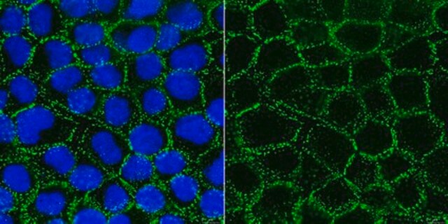
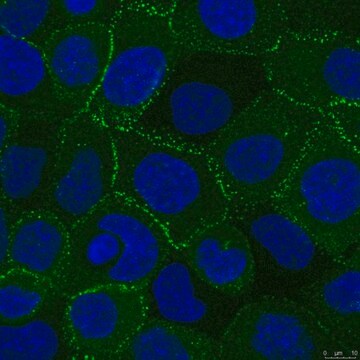
Anti-Desmoglein-1, clone 32-2B detects levels of Desmoglein-1 proteins & has been published & validated for use in WB, IF, IHC & ICC.
Immunohistochemistry Analysis: A 1:1,000 dilution from a representative lot detected Desmoglein-1 in human tonsil tissues.

Immunofluorescence Analysis: A 1:1,000 dilution from a representative lot detected Desmoglein-1 in human tonsil tissues.

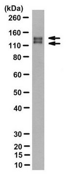
Western Blot Analysis: A representative lot from an independent laboratory detected Desmoglein-1 in bovine epidermal desmosomal cores and in human keratinocyte cell lysates. (Vilela, M. J., et al. (1995). J Cell Sci. 108(Pt 4):1743-1750.).

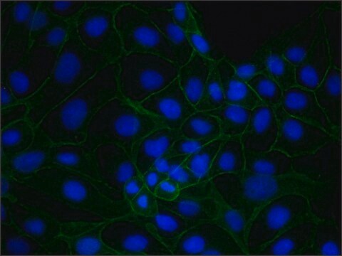
Immunofluorescent Analysis: A representative lot from an independent laboratory detected Desmoglein-1 in MDCK cells (Vilela, M. J., et al. (1995). J Cell Sci. 108(Pt 4):1743-1750.).

Immunohistochemistry Analysis: A representative lot from an independent laboratory detected Desmoglein-1 in human epidermal tissue (Maruani, A., et al. (2008). Am J Clin Pathol. 130(3):369-374.).
Research Category
Cell Structure
Research Sub Category
ECM Proteins

Quality

Evaluated by Western Blot in human tonsil tissue lysate.

Western Blot Analysis: 0.5 µg/mL of this antibody detected Desmoglein-1 in 10 µg of human tonsil tissue lysate.

Target description

~150 kDa observed. This protein may be glycosolated. Uniprot describes a molecular weight at ~112 kDa; however, Desmoglein-1 has been observed at ~150 kDa (Vilela, M. J., et al. (1995). J Cell Sci. 108(Pt 4):1743-1750.).

Physical form

Format: Purified
Protein G Purified
Purified mouse monoclonal IgG2aκ in buffer containing 0.1 M Tris-Glycine (pH 7.4), 150 mM NaCl with 0.05% sodium azide.

Storage and Stability

Stable for 1 year at 2-8°C from date of receipt.

Analysis Note

Control
Human tonsil tissue lysate.

Other Notes

Concentration: Please refer to the Certificate of Analysis for the lot-specific concentration.

Disclaimer

Unless otherwise stated in our catalog or other company documentation accompanying the product(s), our products are intended for research use only and are not to be used for any other purpose, which includes but is not limited to, unauthorized commercial uses, in vitro diagnostic uses, ex vivo or in vivo therapeutic uses or any type of consumption or application to humans or animals.

Not finding the right product?  

Try our Product Selector Tool.

Storage Class Code

12 - Non Combustible Liquids

WGK

WGK 1

Flash Point(F)

Not applicable

Flash Point(C)

Not applicable


Certificates of Analysis (COA)

Search for Certificates of Analysis (COA) by entering the products Lot/Batch Number. Lot and Batch Numbers can be found on a product’s label following the words ‘Lot’ or ‘Batch’.

Already Own This Product?

Find documentation for the products that you have recently purchased in the Document Library.

Visit the Document Library

Immunohistochemical staining with monoclonal antibody 32-2B to desmosomal glycoprotein 1. Its role in the histological assessment of urothelial carcinomas.
Conn, I G, et al.
British Journal of Urology, 65, 176-180 (1990)
Immunohistochemical detection of desmosomes in oral squamous cell carcinomas: correlation with differentiation, mode of invasion, and metastatic potential.
Harada, T, et al.
International Journal of Oral and Maxillofacial Surgery, 21, 346-349 (1992)
Immunostaining with antibodies to desmoglein provides the diagnosis of drug-induced pemphigus and allows prediction of outcome.
Maruani, Annabel, et al.
American Journal of Clinical Pathology, 130, 369-374 (2008)
A simple epithelial cell line (MDCK) shows heterogeneity of desmoglein isoforms, one resembling pemphigus vulgaris antigen.
Vilela, M J, et al.
Journal of Cell Science, 108 ( Pt 4), 1743-1750 (1995)
M J Vilela et al.
The Journal of pathology, 153(4), 365-375 (1987-12-01)
Desmosomes are intercellular adhesive junctions that occur in almost all epithelia and should therefore be useful as epithelial markers in tumour diagnosis. Here, we describe a monoclonal antibody, 32-2B, to a major desmosomal glycoprotein (dgl) which reacts with human tissues

Our team of scientists has experience in all areas of research including Life Science, Material Science, Chemical Synthesis, Chromatography, Analytical and many others.

Contact Technical Service