Skip to Content
Merck
All Photos(2)

Key Documents

07-2167

Sigma-Aldrich

Anti-SUMO 2/3 Antibody

from rabbit, purified by affinity chromatography

Synonym(s):

SMT3 suppressor of mif two 3 homolog 2 (S. cerevisiae), Ubiquitin-like protein SMT3B, SMT3 homolog 2, SMT3 (suppressor of mif two 3, yeast) homolog 2, SMT3 suppressor of mif two 3 homolog 2 (yeast), small ubiquitin-related modifier 2, small ubiquitin-lik

Sign Into View Organizational & Contract Pricing


About This Item

UNSPSC Code:
12352203
eCl@ss:
32160702

biological source

rabbit

Quality Level

antibody product type

primary antibodies

clone

polyclonal

purified by

affinity chromatography

species reactivity

pig, human, mouse, rat

species reactivity (predicted by homology)

bovine (based on 100% sequence homology), porcine (based on 100% sequence homology), rhesus macaque (based on 100% sequence homology), opossum (based on 100% sequence homology), chimpanzee (based on 100% sequence homology)

technique(s)

immunoprecipitation (IP): suitable
western blot: suitable

UniProt accession no.

shipped in

wet ice

target post-translational modification

unmodified

Gene Information

human ... SUMO2(6613)
mouse ... Sumo2(170930)
opossum ... Sumo2(123244757)
rat ... Sumo2(690244)

General description

Small ubiquitin-related modifier 2 (UniProt P61956; also known as HSMT3, Sentrin-2, SMT3 homolog 2, Smt3B, SUMO-2, Ubiquitin-like protein SMT3B) and 3 (UniProt P55854; also known as SMT3 homolog 1, Smt3A, SUMO-3, Ubiquitin-like protein SMT3A) are encoded by the SUMO2 (also known as SMT3B, SMT3H2; Gene ID 6613) and SUMO3 (also known as SMT3A, SMT3H1; Gene ID 6612) genes in human. SUMOylation, protein post-translation modification by small ubiquitin-like modifier (SUMO), is a signaling event in many cellular processes. SUMO proteins are translated as immature precursors and subsequently converted to their mature forms through the activity of sentrin/SUMO-specific proteases (SENPs). SUMOylation is a reversible process. SUMO E1 activating enzyme, E2 conjugating enzyme, and E3 ligase mediate SUMOylation of substrate proteins, while SENPs are responsible for the de-SUMOylation. SUMOylation usually occurs at lysine residues in the consensus KxD/E motif, although not all such lysines become SUMOylated and SUMOylation can also occur on lysine residues outside of this motif. SUMO2 and 3 share 97% identity at the amino acid level, while SUMO1 shares only about 50% sequence homology with SUMO-2 and 3. In addition to difference in their target substrates, SUMO2/3 can be SUMOylated and form chains, whereas SUMO1 cannot and may serve as chain terminator. SUMO-2 can be covalently attached to proteins as a monomer or as a lysine-linked polymer. Its covalent attachment to its substrate via an isopeptide bond requires prior activation by the E1 complex SAE1-SAE2 and linkage to the E2 enzyme UBE2I, and can be promoted by an E3 ligase such as PIAS1-4, RANBP2, CBX4 or ZNF451. This post-translational modification on lysine residues of proteins plays a crucial role in a number of cellular processes such as nuclear transport, DNA replication and repair, mitosis and signal transduction. Although SUMO-2 functions in a manner similar to ubiquitin in that it is bound to target proteins as part of a post-translational modification system, however, unlike ubiquitin which targets proteins for degradation, SUMO-2 is involved in a variety of cellular processes, such as nuclear transport, transcriptional regulation, apoptosis, and protein stability. It is not active until the last two amino acids of the carboxy-terminus (aa 94-95; propeptide) have been cleaved off.

Specificity

This antibody recognizes SUMO 2/3 at the N-terminus.

Immunogen

Epitope: N-terminus
KLH-conjugated linear peptide corresponding to the first 15 amino acids from the N-terminal region of human SUMO-2/3.

Application

Research Category
Signaling
Research Sub Category
Ubiquitin & Ubiquitin Metabolism
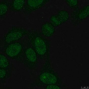
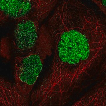
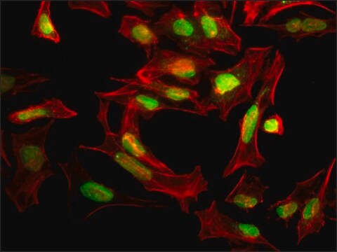
This SUMO 2/3 antibody is validated for use in WB & IP for the detection of the SUMO 2/3 protein.
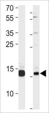
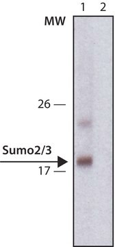
Western Blot (Snap i.d.) Analysis: 5 µg/mL from a previous lot detected SUMO 2/3 on 10 µg of HeLa nuclear extract.
Immunoprecipitation Analysis: A previous was used by an independent laboratory in IP. (Li, T., et al. (2006). The Journal of Biological Chemistry. 281(47):36221-36227.)

Quality

Evaluated by Western Blot in HeLa nuclear extract.
Western Blot Analysis: 1 µg/mL of this antibody detected SUMO 2/3 on 10 µg of HeLa nuclear extract.

Target description

~12 kDa observed; 10.87 for SUMO-2 and 11.64 for SUMO-3 calculated. Uncharacterized bands may be observed in some lysate(s).

Physical form

Affinity purified
Purified rabbit polyclonal antibody in buffer containing 0.1M Tris-Glycine, 0.15M NaCl, 0.05% sodium azide, pH7.4 without azide.

Storage and Stability

Stable for 1 year at 2-8°C from date of receipt.

Analysis Note

Control
HeLa nuclear extract

Other Notes

Concentration: Please refer to the Certificate of Analysis for the lot-specific concentration.

Disclaimer

Unless otherwise stated in our catalog or other company documentation accompanying the product(s), our products are intended for research use only and are not to be used for any other purpose, which includes but is not limited to, unauthorized commercial uses, in vitro diagnostic uses, ex vivo or in vivo therapeutic uses or any type of consumption or application to humans or animals.

Not finding the right product?  

Try our Product Selector Tool.

Storage Class Code

12 - Non Combustible Liquids

WGK

WGK 1

Flash Point(F)

Not applicable

Flash Point(C)

Not applicable


Certificates of Analysis (COA)

Search for Certificates of Analysis (COA) by entering the products Lot/Batch Number. Lot and Batch Numbers can be found on a product’s label following the words ‘Lot’ or ‘Batch’.

Already Own This Product?

Find documentation for the products that you have recently purchased in the Document Library.

Visit the Document Library

Nan Zhao et al.
Nature structural & molecular biology, 25(9), 885-893 (2018-09-05)
Viral infection perturbs host cells and can be used to uncover regulatory mechanisms controlling cellular responses and susceptibility to infections. Using cell biological, biochemical, and genetic tools, we reveal that influenza A virus (IAV) infection induces global transcriptional defects at

Our team of scientists has experience in all areas of research including Life Science, Material Science, Chemical Synthesis, Chromatography, Analytical and many others.

Contact Technical Service