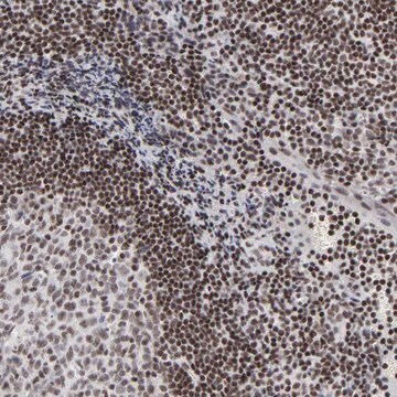
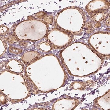
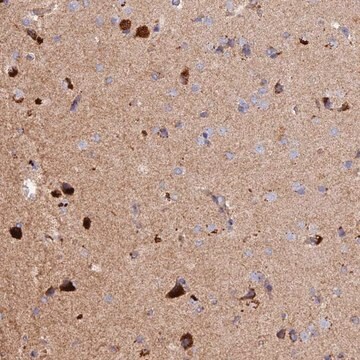
HPA042250
Anti-TADA3 antibody produced in rabbit
Prestige Antibodies® Powered by Atlas Antibodies, affinity isolated antibody, buffered aqueous glycerol solution
别名:
Anti-Ada3, Anti-Flj20221, Anti-Flj21329, Anti-Hada3, Anti-Ngg1, Anti-Tada3l, Anti-Transcriptional adaptor 3
登录查看公司和协议定价
所有图片(4)
About This Item
推荐产品
生物源
rabbit
共軛
unconjugated
抗體表格
affinity isolated antibody
抗體產品種類
primary antibodies
無性繁殖
polyclonal
產品線
Prestige Antibodies® Powered by Atlas Antibodies
形狀
buffered aqueous glycerol solution
物種活性
human, mouse, rat
技術
immunoblotting: 0.04-0.4 μg/mL
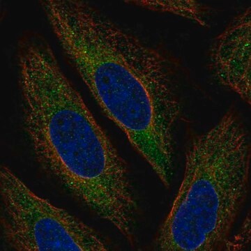
immunofluorescence: 0.25-2 μg/mL
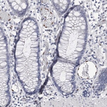
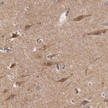
immunohistochemistry: 1:50-1:200
免疫原序列
LAKEEVSRQELRQRVRMADNEVMDAFRKIMAARQKKRTPTKKEKDQAWKTLKERESILKLLDG
UniProt登錄號
運輸包裝
wet ice
儲存溫度
−20°C
目標翻譯後修改
unmodified
基因資訊
human ... TADA3(10474)
免疫原
transcriptional adaptor 3 recombinant protein epitope signature tag (PrEST)
應用
All Prestige Antibodies Powered by Atlas Antibodies are developed and validated by the Human Protein Atlas (HPA) project and as a result, are supported by the most extensive characterization in the industry.
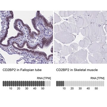
The Human Protein Atlas project can be subdivided into three efforts: Human Tissue Atlas, Cancer Atlas, and Human Cell Atlas. The antibodies that have been generated in support of the Tissue and Cancer Atlas projects have been tested by immunohistochemistry against hundreds of normal and disease tissues and through the recent efforts of the Human Cell Atlas project, many have been characterized by immunofluorescence to map the human proteome not only at the tissue level but now at the subcellular level. These images and the collection of this vast data set can be viewed on the Human Protein Atlas (HPA) site by clicking on the Image Gallery link. We also provide Prestige Antibodies® protocols and other useful information.
The Human Protein Atlas project can be subdivided into three efforts: Human Tissue Atlas, Cancer Atlas, and Human Cell Atlas. The antibodies that have been generated in support of the Tissue and Cancer Atlas projects have been tested by immunohistochemistry against hundreds of normal and disease tissues and through the recent efforts of the Human Cell Atlas project, many have been characterized by immunofluorescence to map the human proteome not only at the tissue level but now at the subcellular level. These images and the collection of this vast data set can be viewed on the Human Protein Atlas (HPA) site by clicking on the Image Gallery link. We also provide Prestige Antibodies® protocols and other useful information.
特點和優勢
Prestige Antibodies® are highly characterized and extensively validated antibodies with the added benefit of all available characterization data for each target being accessible via the Human Protein Atlas portal linked just below the product name at the top of this page. The uniqueness and low cross-reactivity of the Prestige Antibodies® to other proteins are due to a thorough selection of antigen regions, affinity purification, and stringent selection. Prestige antigen controls are available for every corresponding Prestige Antibody and can be found in the linkage section.
Every Prestige Antibody is tested in the following ways:
Every Prestige Antibody is tested in the following ways:
- IHC tissue array of 44 normal human tissues and 20 of the most common cancer type tissues.
- Protein array of 364 human recombinant protein fragments.
聯結
Corresponding Antigen APREST70982
外觀
Solution in phosphate buffered saline, pH 7.2, containing 40% glycerol and 0.02% sodium azide.
法律資訊
Prestige Antibodies is a registered trademark of Merck KGaA, Darmstadt, Germany
免責聲明
Unless otherwise stated in our catalog or other company documentation accompanying the product(s), our products are intended for research use only and are not to be used for any other purpose, which includes but is not limited to, unauthorized commercial uses, in vitro diagnostic uses, ex vivo or in vivo therapeutic uses or any type of consumption or application to humans or animals.
未找到合适的产品?
试试我们的产品选型工具.
儲存類別代碼
10 - Combustible liquids
水污染物質分類(WGK)
WGK 1
閃點(°F)
Not applicable
閃點(°C)
Not applicable
Nikolaos Vosnakis et al.
The EMBO journal, 36(18), 2710-2725 (2017-07-21)
SAGA and ATAC are two distinct chromatin modifying co-activator complexes with distinct enzymatic activities involved in RNA polymerase II (Pol II) transcription regulation. To investigate the mobility of co-activator complexes and general transcription factors in live-cell nuclei, we performed imaging
Wenyi Mi et al.
Nature communications, 9(1), 3759-3759 (2018-09-16)
Recognition of histones by epigenetic readers is a fundamental mechanism for the regulation of chromatin and transcription. Most reader modules target specific post-translational modifications on histones. Here, we report the identification of a reader of histone H3, the ZZ-type zinc
Wenyi Mi et al.
Nature communications, 8(1), 1088-1088 (2017-10-24)
Recognition of modified histones by "reader" proteins constitutes a key mechanism regulating diverse chromatin-associated processes important for normal and neoplastic development. We recently identified the YEATS domain as a novel acetyllysine-binding module; however, the functional importance of YEATS domain-containing proteins
Shashank Srivastava et al.
Cell cycle (Georgetown, Tex.), 16(16), 1515-1525 (2017-08-02)
The ADA3 (Alteration/Deficiency in Activation 3) protein is an essential adaptor component of several Lysine Acetyltransferase (KAT) complexes involved in chromatin modifications. Previously, we and others have demonstrated a crucial role of ADA3 in cell cycle progression and in maintenance
Shakur Mohibi et al.
The Journal of biological chemistry, 290(47), 28299-28310 (2015-10-03)
ADA3 (alteration/deficiency in activation 3) is a conserved component of several transcriptional co-activator and histone acetyltransferase (HAT) complexes. Recently, we generated Ada3 knock-out mice and demonstrated that deletion of Ada3 leads to early embryonic lethality. The use of Ada3(FL/FL) mouse
我们的科学家团队拥有各种研究领域经验,包括生命科学、材料科学、化学合成、色谱、分析及许多其他领域.
联系技术服务部门
