一般說明
Intermediate-sized filaments are abundant cytoplasmic structural proteins in most vertebrate cells. Cytokeratins, a group of at least 29 different proteins, are characteristic of epithelial and trichocytic cells. Cytokeratins 4, 5, 6 and 8 are members of the type II neutral-to-basic subfamily. Cytokeratin peptide 4 (59 kDa) is the secondary type II keratin expressed in noncornified stratified squamous epithelia. Cytokeratin peptide 5 (58 kDa) is the primary type II keratin in stratified epithelia, while cytokeratin type 8 (52 kDa) is a major type II keratin in simple epithelia. Cytokeratin 6 (56 kDa) is a "hyperproliferation" cytokeratin expressed in tissues with natural or pathological high turnover. Cytokeratins 10, 13 and 18 are members of the type I acidic subfamily. Cytokeratin peptide 10 (56 kDa) is the secondary type I keratin expressed in cornified epithelia. Cytokeratin 13 (54 kDa) is the secondary type I keratin expressed in non-cornified stratified squamous epithelia. Cytokeratin 18 (45 kDa) is the primary type I keratin expressed in simple epithelial cells.
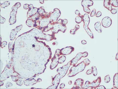
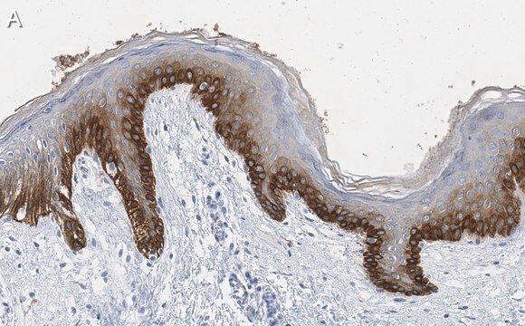
Monoclonal Anti-Pan Cytokeratin (clone C-11) recognizes human cytokeratins 4, 5, 6, 8, 10, 13 and 18 in immunoblotting. The antibody reacts with simple, cornifying and non-cornifying squamous epithelia and pseudostratified epithelia. It does not react with non-epithelial normal human tissues. This antibody can be applied to methanol or acetone-fixed, frozen sections, and to formalin-fixed, paraffin-embedded human tissues. Increased staining intensity is seen following proteolytic treatment of formalin fixed tissue. Similarly, methacarn-fixed material is also suitable for cytokeratin demonstration. Monoclonal Anti-Pan cytokeratin exhibits a wide interspecies cross-reactivity (e.g., human, bovine, rat, frog).
免疫原
keratin-enriched preparation from cultured human epidermoid carcinoma cell line A431.
應用
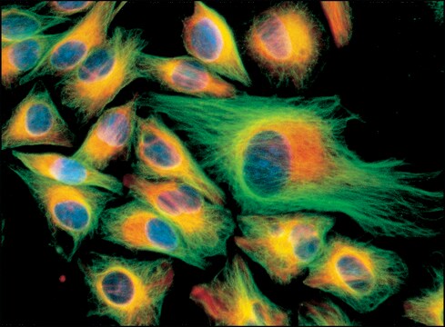
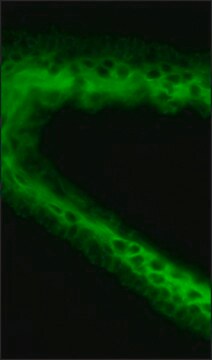
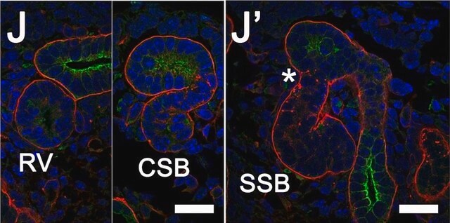
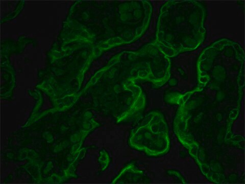
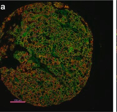
Monoclonal Anti-Cytokeratin, pan antibody has been used in immunofluorescence microscopy.
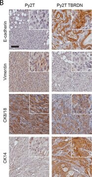
Monoclonal anti-cytokeratins are specific markers of epithelial cell differentiation and have been widely used as tools in tumor identification and classification. Mouse monoclonal clone C-11 anti-cytokeratin, pan antibody is a broad spectrum antibody which recognizes an epitope present in most human epithelial tissues. It facilitates typing of normal, metaplastic and neoplastic cells. It may aid in the discrimination of carcinomas and non-epithelial tumors such as sarcomas, lymphomas and neural tumors. It is also useful in detecting micrometastases in lymph nodes, bone marrow and other tissues, and for determining the origin of poorly differentiated tumors. It is useful for staining of cultured epithelial cell lines. Mouse monoclonal clone C-11 anti-Cytokeratin, pan antibody may be used for the localization of cytokeratins using various immunochemical assays such as immunoblotting, dot blotting and immunohistochemistry (immunofluorescence and immunoenzymatic staining).
生化/生理作用
Cytokeratins are usually used to determine epithelial differentiation. They help to maintain the integrity of the epithelium of the anterior segment of the eye.
免責聲明
Unless otherwise stated in our catalog or other company documentation accompanying the product(s), our products are intended for research use only and are not to be used for any other purpose, which includes but is not limited to, unauthorized commercial uses, in vitro diagnostic uses, ex vivo or in vivo therapeutic uses or any type of consumption or application to humans or animals.

