跳转至内容
Merck

B6529

Sigma-Aldrich

亮蓝R染色液

ethanol solution

别名:

酸性蓝83, Coomassie 亮蓝R, 亮吲哚蓝蛋白 6B, 酸性蓝 83

登录查看公司和协议定价


About This Item

经验公式(希尔记法):
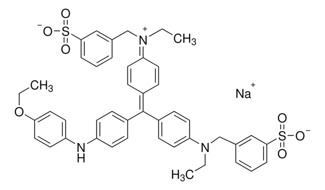
C45H44N3NaO7S2
CAS号:
分子量:
825.97
顏色索引編號:
42660
Beilstein:
5718025
MDL號碼:
分類程式碼代碼:
12171500
PubChem物質ID:
NACRES:
NA.47

product name

亮蓝R染色液, suitable for (for immunoelectrophoresis protein staining)

形狀

liquid

品質等級

顏色

dark blue

適合性

suitable for (for immunoelectrophoresis protein staining)

應用

diagnostic assay manufacturing
hematology
histology

儲存溫度

room temp

SMILES 字串

[Na+].CCOc1ccc(Nc2ccc(cc2)C(\c3ccc(cc3)N(CC)Cc4cccc(c4)S([O-])(=O)=O)=C5\C=C/C(C=C5)=[N+](\CC)Cc6cccc(c6)S([O-])(=O)=O)cc1

InChI

1S/C45H45N3O7S2.Na/c1-4-47(31-33-9-7-11-43(29-33)56(49,50)51)40-23-15-36(16-24-40)45(35-13-19-38(20-14-35)46-39-21-27-42(28-22-39)55-6-3)37-17-25-41(26-18-37)48(5-2)32-34-10-8-12-44(30-34)57(52,53)54;/h7-30H,4-6,31-32H2,1-3H3,(H2,49,50,51,52,53,54);/q;+1/p-1

InChI 密鑰

NKLPQNGYXWVELD-UHFFFAOYSA-M

正在寻找类似产品? 访问 产品对比指南

應用

亮蓝R染色溶液专门设计用于在免疫电泳和Ouchterlony凝胶之后对琼脂糖凝胶中的蛋白质进行染色。污渍含有乙醇和乙酸,因此在染色前无需固定凝胶。首先用水和盐水洗涤免疫电泳或Ouchterlony凝胶,以去除未沉淀的蛋白质,然后干燥。然后将凝胶浸入染色溶液中30分钟,并用10%乙酸脱色。
电泳后检测蛋白条带。

成分

0.5% (w/v) 亮蓝 R、45% (v/v) 乙醇和 10% (v/v) 乙酸。

分析報告

在免疫电泳后测试琼脂糖凝胶的适用性。

法律資訊

象形圖

FlameExclamation mark

訊號詞

Warning

危險聲明

危險分類

Eye Irrit. 2 - Flam. Liq. 3 - Skin Irrit. 2

儲存類別代碼

3 - Flammable liquids

水污染物質分類(WGK)

WGK 2

閃點(°F)

81.0 °F - closed cup

閃點(°C)

27.2 °C - closed cup

個人防護裝備

Faceshields, Gloves, Goggles, type ABEK (EN14387) respirator filter


从最新的版本中选择一种:

分析证书(COA)

Lot/Batch Number

没有发现合适的版本?

如果您需要特殊版本,可通过批号或批次号查找具体证书。

已有该产品?

在文件库中查找您最近购买产品的文档。

访问文档库

Wei Zhang et al.
International journal of oncology, 54(5), 1719-1733 (2019-03-14)
Ovarian cancer remains the most lethal type of cancer among all gynecological malignancies. The majority of patients are diagnosed with ovarian cancer at the late stages of the disease. Therefore, there exists an imperative need for the development of early
K Kubo
Analytical biochemistry, 213(2), 200-205 (1993-09-01)
In sodium dodecyl sulfate-polyacrylamide gel electrophoresis (SDS-PAGE) according to the method of Weber and Osborn, protein bands are often distorted by tailing at both ends, and the phenomenon is often called "edge tailing." This was eliminated by adding glycerol at
Zhang Xin-Guo Chen Jian-Hua et al.
Journal of chromatographic science, 50(9), 820-825 (2012-06-22)
A human serum albumin and Thymosin α1 (HSA-Tα1) fusion protein was designed and over-expressed in Pichia pastoris. To purify the fusion protein, a new native preparative electrophoresis system that involved a modified device with a sample receiving chamber, and an
Reiner Westermeier
Proteomics, 6 Suppl 2, 61-64 (2006-10-13)
In spite of the high sensitivity of silver staining and the wide dynamic range of various fluorescent detection methods, Coomassie Brilliant Blue staining is still the most widely used protein detection technique for proteins separated by polyacrylamide gel electrophoresis. There
Chun Yi Liau et al.
Journal of bioscience and bioengineering, 106(1), 111-113 (2008-08-12)
A modified Coomassie Brilliant Blue G 250 staining method for detecting chitinolytic enzymes in chitin-containing polyacrylamide gel electrophoresis (PAGE) is presented. The staining formed achromatic zones at the locations of the migrated enzyme. Using Streptomyces griseus chitinase, we have demonstrated

商品

MISSION® Target ID Library for Human miRNA Target Identification and Discovery;

To meet the great diversity of protein analysis needs, Sigma offers a wide selection of protein visualization (staining) reagents. EZBlue™ and ProteoSilver™, designed specifically for proteomics, also perform impressively in traditional PAGE formats.

我们的科学家团队拥有各种研究领域经验,包括生命科学、材料科学、化学合成、色谱、分析及许多其他领域.

联系技术服务部门