推荐产品
生物源
mouse
品質等級
抗體表格
purified immunoglobulin
抗體產品種類
primary antibodies
無性繁殖
12D10, monoclonal
物種活性
human
物種活性(以同源性預測)
all
技術
flow cytometry: suitable
immunocytochemistry: suitable
immunofluorescence: suitable
immunohistochemistry: suitable
immunoprecipitation (IP): suitable
western blot: suitable
同型
IgG2aκ
運輸包裝
wet ice
目標翻譯後修改
unmodified
基因資訊
human ... NPEPPS(9520)
一般說明
嘌呤霉素是一种氨基核苷酸抗生素,来源于白黑链霉菌,是蛋白合成抑制剂,可通过核糖体中的过早链终止来阻止翻译。抗嘌呤霉素的单克隆抗体可与标准免疫化学方法一起用于直接监测翻译,这种方法称为翻译表面感应(SUnSET)。该分子的一部分类似于氨基酰化tRNA的3′端,这使其可用于蛋白质翻译分析。嘌呤霉素诱导胸腺细胞和HL-60白血病细胞内DNA断裂。
特異性
已证明抗嘌呤霉素抗体(克隆号12D10)可与嘌呤霉素预孵育的人类检测样品反应。当检测样品与嘌呤霉素一起孵育时,预计会与所有物种发生反应。
免疫原
来源于白黑链霉菌的嘌呤霉素
應用
抗嘌呤霉素抗体,克隆 12D10,可检测掺入到蛋白质中的嘌呤霉素。抗嘌呤霉素的单克隆抗体可与标准免疫化学方法一起使用。
研究子范畴
RNA代谢&结合蛋白
RNA代谢&结合蛋白
研究类别
表观遗传学&核功能
表观遗传学&核功能
蛋白印迹分析(总蛋白染色):使用SDS-PAGE分离用嘌呤霉素和环己酰胺处理或仅用嘌呤霉素处理的HEK293细胞裂解液,然后转移到膜上。采用Ponceau S染色法观察蛋白质。
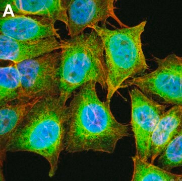
免疫细胞化学分析:代表批次的1:10,000稀释液可在用嘌呤霉素处理的HeLa细胞中检测到掺入嘌呤霉素的新合成蛋白。
蛋白印迹分析:A representative lot detected Puromycin-incorporated neosynthesized proteins in WB (Reineke, L. C., et al. (2012).Mol Biol Cell.23(18):3499-3510.; Trinh, M. A. et al. (2012).Cell Rep.1(6):678-688.; Fortin, D. A., et al. (2012).J Neurosci. 32(24):8127-8137.;Clavarino, G., et al. (2012).PLoS Pathog.8(5):e1002708.;David, A., et al. (2012).J Cell Biol. 197(1):45-57.;White, L. K., et al. (2011).J Virol.85(1):606-620.; Hoeffer, C. A., et al. (2011).Proc Natl Acad Sci USA.108(8):3383-3388.;Goodman, C. A., et al. (2010).FASEB J. 25(3):1028-1039.;Schmidt, E., K., et al. (2009).Nat Methods.6(4):275-277.;Santini, E., et al. (2013).Nature.493(7432):411-415.; Quy.P. N., et al. (2013).J Biol Chem. 288(2):1125-1134.;Hulmi.J. J., et al. (2012).Am J Physiol Endocrinol Metab.304(1):E41-50.;Bhattacharya, A., et al. (2012).Neuron.76(2):325-337.; Hoeffer, C. A., et al. (2013).J Neurophysiol.109(1):68-76.).
Immunofluorescence Analysis: A representative lot detected Puromycin-incorporated neosynthesized proteins in WB (Reineke, L. C., et al. (2012).Mol Biol Cell.23(18):3499-3510.; Trinh, M. A. et al. (2012).Cell Rep.1(6):678-688.; Fortin, D. A., et al. (2012).J Neurosci. 32(24):8127-8137.;David, A., et al. (2012).J Cell Biol. 197(1):45-57.;David, A., et al. (2011).J Biol Chem. 286(23):20688-20700.;White, L. K., et al. (2011).J Virol.85(1):606-620.; Hoeffer, C. A., et al. (2011).Proc Natl Acad Sci USA.108(8):3383-3388.;Schmidt, E., K., et al. (2009).Nat Methods.6(4):275-277.; Goodman, C. A., et al. (2012).Proc Natl Acad Sci USA.109(17):E989.; Santini, E., et al. (2013).Nature.493(7432):411-415.; Quy.P. N., et al. (2013).J Biol Chem. 288(2):1125-1134.).
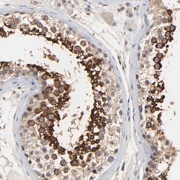
Immunohistochemistry Analysis: A representative lot detecte Puromycin-incorporated neosynthesized protein in IHC (Goodman, C. A., et al. (2010).FASEB J. 25(3):1028-1039.).
Fluorescence Activated Cell Sorting Analysis: A representative lot detected Puromycin-incorporated neosynthesized proteins in FACS (David, A., et al. (2012).J Cell Biol. 197(1):45-57.; Schmidt, E., K., et al. (2009).Nat Methods.6(4):275-277.)。
Alexa Fluor™是Life Technologies的注册商标。
品質
通过对用嘌呤霉素和环己酰胺处理或仅用嘌呤霉素处理的HEK293细胞裂解物进行蛋白印迹分析而进行评估。
蛋白印迹分析:该抗体的1:25,000稀释液可在仅用嘌呤霉素处理的HEK293细胞裂解液中检测到掺入嘌呤霉素的新合成蛋白。该抗体还在用嘌呤霉素和环己酰胺处理的HEK293细胞中检测到少量的掺入嘌呤霉素的新合成蛋白。
蛋白印迹分析:该抗体的1:25,000稀释液可在仅用嘌呤霉素处理的HEK293细胞裂解液中检测到掺入嘌呤霉素的新合成蛋白。该抗体还在用嘌呤霉素和环己酰胺处理的HEK293细胞中检测到少量的掺入嘌呤霉素的新合成蛋白。
標靶描述
将嘌呤霉素掺入新合成蛋白中。在仅存在嘌呤霉素的情况下,该抗体可检测到掺入嘌呤霉素的多种分子量的新合成蛋白。但是,在同时存在真核生物蛋白质生物合成抑制剂环己酰胺的情况下,观察到的信号较弱。
外觀
形式:纯化
纯化的小鼠单克隆IgG2aκ,溶于含有0.1 M Tris-甘氨酸(pH 7.4,150 mM NaCl)和0.05%叠氮化钠的缓冲液中。
蛋白G纯化
儲存和穩定性
自发运之日起,在 2-8°C 条件下可稳定保存1年
分析報告
对照
用嘌呤霉素和环己酰胺处理或仅用嘌呤霉素处理的HEK293细胞裂解物。
用嘌呤霉素和环己酰胺处理或仅用嘌呤霉素处理的HEK293细胞裂解物。
其他說明
浓度:请参考批次特异性浓缩物的分析证书。
法律資訊
ALEXA FLUOR is a trademark of Life Technologies
免責聲明
除非我们的产品目录或产品附带的其他公司文档另有说明,否则我们的产品仅供研究使用,不得用于任何其他目的,包括但不限于未经授权的商业用途、体外诊断用途、离体或体内治疗用途或任何类型的消费或应用于人类或动物。
未找到合适的产品?
试试我们的产品选型工具.
儲存類別代碼
12 - Non Combustible Liquids
水污染物質分類(WGK)
WGK 1
閃點(°F)
Not applicable
閃點(°C)
Not applicable
Alexandre David et al.
The Journal of cell biology, 197(1), 45-57 (2012-04-05)
Whether protein translation occurs in the nucleus is contentious. To address this question, we developed the ribopuromycylation method (RPM), which visualizes translation in cells via standard immunofluorescence microscopy. The RPM is based on ribosome-catalyzed puromycylation of nascent chains immobilized on
Chikungunya virus induces IPS-1-dependent innate immune activation and protein kinase R-independent translational shutoff.
White, LK; Sali, T; Alvarado, D; Gatti, E; Pierre, P; Streblow, D; Defilippis, VR
Journal of virology null
SUnSET, a nonradioactive method to monitor protein synthesis.
Schmidt, Enrico K, et al.
Nature Methods, 6, 275-277 (2009)
Imaging of protein synthesis with puromycin.
Goodman, Craig A, et al.
Proceedings of the National Academy of Sciences of the USA, 109, E989-E989 (2012)
Craig A Goodman et al.
FASEB journal : official publication of the Federation of American Societies for Experimental Biology, 25(3), 1028-1039 (2010-12-15)
In this study, the principles of surface sensing of translation (SUnSET) were used to develop a nonradioactive method for ex vivo and in vivo measurements of protein synthesis (PS). Compared with controls, we first demonstrate excellent agreement between SUnSET and
我们的科学家团队拥有各种研究领域经验,包括生命科学、材料科学、化学合成、色谱、分析及许多其他领域.
联系技术服务部门


