推荐产品
生物源
rat
品質等級
抗體表格
purified antibody
抗體產品種類
primary antibodies
無性繁殖
1D4, monoclonal
物種活性
human
物種活性(以同源性預測)
mouse (based on 100% sequence homology)
技術
flow cytometry: suitable
immunocytochemistry: suitable
immunohistochemistry: suitable
western blot: suitable
同型
IgG2aκ
NCBI登錄號
UniProt登錄號
運輸包裝
wet ice
目標翻譯後修改
unmodified
基因資訊
human ... PLET1(349633)
一般說明
Placenta-expressed transcript-1 (Plet-1) is a recently identified protein that as of yet has no known function and is not associated with any one family of proteins. Its observation has been restricted to developing mesonephros and pharyngeal endoderm. Observation in adult tissue is more widely expressed, having been found in several locations such as mammary, pancreas and prostate epithelia. Its location in the pancreas is notable as the pancreas is a well known reserve for stem cells and progenitor cells. Within the pancreas, Plet-1 has been shown to be localized in the major duct epithelium, and as such will serve as an excellent marker for epithelial progenitor cells and stem cells, and as a useful implement for observing the genetics of linear relationships and the operation of molecular mechanisms for homeostasis, development and wound healing in organ and tissue systems.
Recently, Plet-1 has been identified as a specific marker of early thymic epithelial progenitor cells. In the postgastrulation embryo, Plet-1 expression is highly restricted to the developing pharyngeal endoderm and mesonephros. This antibody recognizes the same protein as MTS20 and MTS24.
Recently, Plet-1 has been identified as a specific marker of early thymic epithelial progenitor cells. In the postgastrulation embryo, Plet-1 expression is highly restricted to the developing pharyngeal endoderm and mesonephros. This antibody recognizes the same protein as MTS20 and MTS24.
特異性
This antibody recognizes Plet-1.
免疫原
Epitope: Unknown
Full length recombinant protein corresponding to Plet-1.
應用
Flow Cytometry Analysis: A previous lot was used by an independent laboratory in FC (Depreter, M.G.L., et al., 2008).
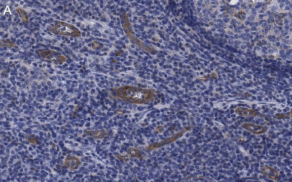
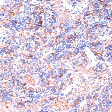
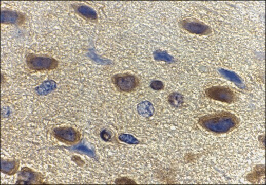
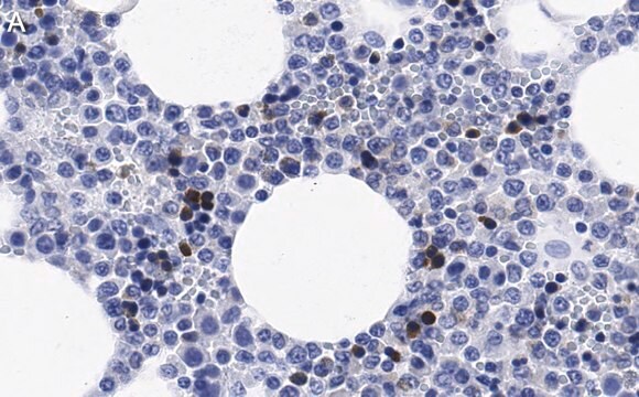
Immunohistochemistry Analysis: A previous lot was used by an independent laboratory in IH (Depreter, M.G.L., et al., 2008).
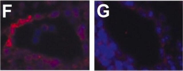
Immunocytochemistry Analysis: A previous lot was used by an independent laboratory in IC (Depreter, M.G.L., et al., 2008).
Immunohistochemistry Analysis: A previous lot was used by an independent laboratory in IH (Depreter, M.G.L., et al., 2008).
Immunocytochemistry Analysis: A previous lot was used by an independent laboratory in IC (Depreter, M.G.L., et al., 2008).
Research Category
Stem Cell Research
Stem Cell Research
Research Sub Category
Pluripotent & Early Differentiation
Pluripotent & Early Differentiation
This Anti-Plet-1 (Placenta-expressed transcript 1 protein) Antibody, clone 1D4 is validated for use in WB, IC, IH, FC for the detection of Plet-1 (Placenta-expressed transcript 1 protein).
品質
Evaluated by Western Blot in human placenta cell lysate.
Western Blot Analysis: A 1:2,500 dilution of this antibody detected Plet-1 in 10 µg of human placenta cell lysate.
Western Blot Analysis: A 1:2,500 dilution of this antibody detected Plet-1 in 10 µg of human placenta cell lysate.
標靶描述
~ 23 kDa
外觀
Protein G Purified
Format: Purified
Purified rat monoclonal IgG2aκ in buffer containing 0.1 M Tris-Glycine (pH 7.4, 150 mM NaCl) with 0.05% sodium azide.
儲存和穩定性
Stable for 1 year at 2-8°C from date of receipt.
分析報告
Control
Human placenta cell lysate
Human placenta cell lysate
免責聲明
Unless otherwise stated in our catalog or other company documentation accompanying the product(s), our products are intended for research use only and are not to be used for any other purpose, which includes but is not limited to, unauthorized commercial uses, in vitro diagnostic uses, ex vivo or in vivo therapeutic uses or any type of consumption or application to humans or animals.
未找到合适的产品?
试试我们的产品选型工具.
儲存類別代碼
12 - Non Combustible Liquids
水污染物質分類(WGK)
WGK 1
閃點(°F)
Not applicable
閃點(°C)
Not applicable
Bénédicte Oulès et al.
Nature communications, 11(1), 5067-5067 (2020-10-22)
Although acne is the most common human inflammatory skin disease, its pathogenic mechanisms remain incompletely understood. Here we show that GATA6, which is expressed in the upper pilosebaceous unit of normal human skin, is down-regulated in acne. GATA6 controls keratinocyte
我们的科学家团队拥有各种研究领域经验,包括生命科学、材料科学、化学合成、色谱、分析及许多其他领域.
联系技术服务部门
