推荐产品
生物源
mouse
抗體表格
purified immunoglobulin
抗體產品種類
primary antibodies
無性繁殖
QBEnd/10, monoclonal
物種活性
human, monkey
包裝
antibody small pack of 25 μL
技術
electron microscopy: suitable
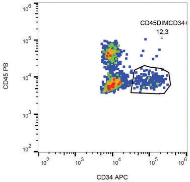
flow cytometry: suitable
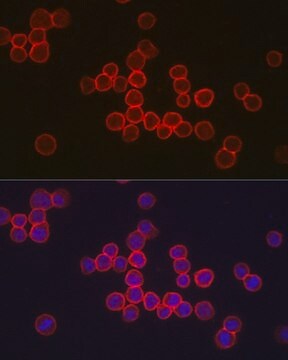
immunofluorescence: suitable
immunohistochemistry: suitable (paraffin)
western blot: suitable
同型
IgG1λ
NCBI登錄號
UniProt登錄號
目標翻譯後修改
unmodified
基因資訊
human ... CD34(947)
一般說明
Hematopoietic progenitor cell antigen CD34 (UniProt: P28906; also known as CD34) is encoded by the CD34 gene (Gene ID: 947) in human. CD34 is a highly glycosylated single-pass type I membrane protein that is expressed on hematopoietic progenitor cells and small vessel endothelium of a variety of tissues. Under normal conditions, CD34+ expressing cells account for about 1 2% of the total bone marrow cells. It serves as an adhesion molecule that plays a role in early hematopoiesis by mediating the attachment of stem cells to the bone marrow extracellular matrix or directly to stromal cells. It is also reported to act as a scaffold for the attachment of lineage specific glycans, allowing stem cells to bind to lectins expressed by stromal cells or other marrow components. CD34 is synthesized with a signal peptide (aa 1-31) that is cleaved off in the mature form. The mature form has an extracellular domain (aa 32-290), a transmembrane domain (aa 291-311), and a cytoplasmic domain (aa 312-385). Two isoforms of CD34 have been described that are produced by alternative splicing.
特異性
Clone QBEnd/10 specifically detects CD34 in human and non-human primates.
免疫原
Human placental endothelial membrane vesicles.
應用
Anti-CD34, clone QBEnd/10, Cat. No. CBL496-I, is a mouse monoclonal antibody that detects CD34 and has been tested for use in Electron Microscopy, Flow Cytometry, Immunofluorescence and Fluorescence Activated Cell Sorting (FACS), Immunohistochemistry (Paraffin), and Western Blotting.
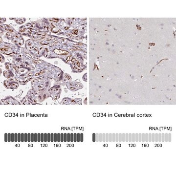
Immunohistochemistry (Paraffin) Analysis: A 1:250 dilution from a representative lot detected CD34 in human brain tissue sections.
Fluorescence Activated Cell Sorting (FACS) Analysis: A representative lot was used to sort CD34+ cells from bone marrow. (de Bock, C.E., et. al. (2012). Leukemia. 26(5):918-26).
Immunofluorescence Analysis: A representative lot detected CD34 in Immunofluorescence applications (Miki, T., et. al. (2010). Mol Cancer Res. 8(5):665-76).
Electron Microscopy Analysis: A representative lot detected CD34 in Electron Microscopy applications (Fina, L., et. al. (1990). Blood. 75(12):2417-26).
Immunohistochemistry Analysis: A representative lot detected CD34 in Immunohistochemistry applications (Engler, J.R., et. al. (2012). PLoS One. 7(8):e43339; Fina, L., et. al. (1990). Blood. 75(12):2417-26).
Flow Cytometry Analysis: A representative lot detected CD34 in Flow Cytometry applications (de Bock, C.E., et. al. (2012). Leukemia. 26(5):918-26; Fina, L., et. al. (1990). Blood. 75(12):2417-26).
Western Blotting Analysis: A representative lot detected CD34 in Western Blotting applications (Fina, L., et. al. (1990). Blood. 75(12):2417-26).
品質
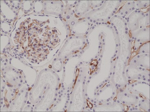
Evaluated by Immunohistochemistry (Paraffin) in human kidney tissue sections.
Immunohistochemistry (Paraffin) Analysis: A 1:250 dilution of this antibody detected CD34 in human kidney tissue sections.
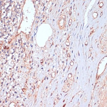
Immunohistochemistry (Paraffin) Analysis: A 1:250 dilution of this antibody detected CD34 in human kidney tissue sections.
標靶描述
40.72 kDa Calculated. This antibody recognizes a heavily glycosylated transmembrane protein: gp 105-120 kDa
外觀
Format: Purified
其他說明
Concentration: Please refer to lot specific datasheet.
未找到合适的产品?
试试我们的产品选型工具.
我们的科学家团队拥有各种研究领域经验,包括生命科学、材料科学、化学合成、色谱、分析及许多其他领域.
联系技术服务部门