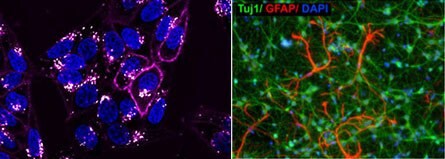
成像分析和活细胞成像

利用活细胞成像和分析技术,科学家可以对动态的细胞事件进行实时研究,从而获得独特的生物学见解。这些技术与PCR、流式细胞术、免疫细胞化学及抗体组织染色等技术不同,后者仅对细胞事件的某一瞬间进行分析。如今,随着相机、活细胞荧光染料、荧光蛋白、视频压缩和延时显微镜等领域的不断成熟,我们得以对活细胞、活细胞间相互作用以及亚细胞过程进行可视化和分析,并获得出色的细节信息和保真度。
相关技术文章
- Cell based assays for cell proliferation (BrdU, MTT, WST1), cell viability and cytotoxicity experiments for applications in cancer, neuroscience and stem cell research.
- Firefly luciferase is a widely used bioluminescent reporter for studying gene regulation and function. It is a very sensitive genetic reporter due to the absence of endogenous luciferase activity in mammalian cells or tissues.
- PKH and CellVue® Fluorescent Cell Linker Kits provide fluorescent labeling of live cells over an extended period of time, with no apparent toxic effects.
- Fluorescent calcium indicators and dyes to measure Ca+ flux used in calcium imaging experiments. Calcium ions (Ca2+) play vital cellular physiology roles in signal transduction pathways, in neurotransmitter release, in contraction of all muscle cell types, as enzyme cofactors, and in fertilization.
- Lipophilic cell tracking dyes enable cancer biologists to track tumor and immune cell functions both in vitro and in vivo. Read the article to choose a right membrane dye kit for cell tracking and proliferation monitoring.
- 查看完整内容 (0)
相关实验方案
- PKH dyes have been successfully used to label exosomes and extracellular vesicles for in vitro and in vivo tracking experiments. The exosome labeling protocol below details steps for reliable fluorescent labeling of exosomes using PKH dyes for microvesicle tracking experiments.
- Cell staining can be divided into four steps: cell preparation, fixation, application of antibody, and evaluation.
- Immunocytochemistry (ICC) Cell Capture Imaging Reagent and protocol for eliminating wash steps, and is highly effective in applications involving rare cells, low cell count samples, and limited access samples.
- This is a Toluidine Blue Staining protocol listing materials and methods.
- This protocol covers 3 modes for the microscopic examination of cell samples.
- 查看完整内容 (0)
活细胞成像系统
许多技术已被用于对复杂的结构和细胞行为进行可视化。在静态分析中,细胞会被固定并染色,这就需要杀死细胞,从而捕获细胞时间中的单个瞬间。尽管静态技术适用于某些应用,但它们并不能提供关于细胞活动的有效信息。利用动态的活细胞方法可以对活细胞中的结构、行为或组成进行可视化,而无需传统的染色。这些技术非常适合必须模拟体内条件的应用,以及开发用于药物筛选的预测性分析。
活细胞成像系统采用先进的光学系统、环境控制和试剂,可实现对哺乳动物、细菌和酵母细胞进行长期的研究。额外采用了微流体技术的培养和成像系统还能实现对培养微环境进行精确的控制,如流量、气体混合物和温度,并同时限制对细胞的物理压力。活细胞成像和分析适用于多种应用,包括缺氧、细胞迁移、3D细胞培养、细胞骨架动力学和蛋白质运输等。先进的系统支持自动化设置培养条件、处理条件的施用和撤销以及图片生成的时间间隔,以静态图片或视频数据的形式呈现。
活细胞成像试剂
活细胞成像培养基和补剂可在延时实验中保护细胞免受光诱导的细胞损伤。这些试剂还经过特殊配制,具有低自发荧光和光致漂白性,从而大大提高了荧光活细胞图像的质量。
我们还可提供大量的活细胞染料,帮助研究活细胞的动态过程。活细胞荧光细胞器染料可对特定细胞器进行选择性染色,如细胞膜、细胞核、细胞质、线粒体、溶酶体、内质网(ER)、高尔基体和细胞骨架蛋白,且不会增加细胞毒性。在活细胞成像中使用细胞器染料作为复染剂,也可在功能研究中发挥作用。
带有GFP和RFP的生物传感器可用于通过荧光显微镜或延时视频捕获来检测特定蛋白质及其在活细胞内的亚细胞定位。慢病毒载体系统是一种将基因和基因产物引入细胞的常用工具,其相比于基于化学的转染具有多种优势。除细胞器检测外,可渗透细胞的活细胞染料还可用于细胞凋亡检测、细胞活力、细胞健康、缺氧、活性氧种类研究、钙指示剂成像以及神经或干细胞实验等应用。
如要继续阅读,请登录或创建帐户。
暂无帐户?