Skip to Content
Merck
All Photos(8)

Key Documents

HPA010136

Sigma-Aldrich

Anti-POR antibody produced in rabbit

enhanced validation

Prestige Antibodies® Powered by Atlas Antibodies, affinity isolated antibody, buffered aqueous glycerol solution

Synonym(s):

Anti-CPR antibody produced in rabbit, Anti-NADPH--cytochrome P450 reductase antibody produced in rabbit, Anti-P450R antibody produced in rabbit

Sign Into View Organizational & Contract Pricing

Select a Size


Select a Size

Change View

About This Item

UNSPSC Code:
12352203
Human Protein Atlas Number:
NACRES:
NA.41

biological source

rabbit

Quality Level

conjugate

unconjugated

antibody form

affinity isolated antibody

antibody product type

primary antibodies

clone

polyclonal

product line

Prestige Antibodies® Powered by Atlas Antibodies

form

buffered aqueous glycerol solution

species reactivity

human, rat, mouse

enhanced validation

orthogonal RNAseq
Learn more about Antibody Enhanced Validation

General description

Cytochrome p450 oxidoreductase (POR) is a 78 kDa membrane-bound diflavin oxidoreductase, which is located on the cytoplasmic side of the endoplasmic reticulum and the outer membrane of the nuclear envelope. The gene is located on human chromosome 7q11.23. POR consists of flavin mononucleotide (FMN) binding domain and nicotinamide adenine dinucleotide phosphate (NADPH)/flavin adenine dinucleotide (FAD) binding domain.
POR (P450 oxidoreductase) is an 80kDa membrane-bound enzyme, localized to endoplasmic reticulum (ER). It is a key enzyme in the P450-mon-oxygenase drug-metabolizing system. It belongs to the diflavin reductases.

Immunogen

NADPH--cytochrome P450 reductase recombinant protein epitope signature tag (PrEST)

Application

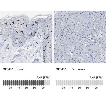
All Prestige Antibodies Powered by Atlas Antibodies are developed and validated by the Human Protein Atlas (HPA) project and as a result, are supported by the most extensive characterization in the industry.

The Human Protein Atlas project can be subdivided into three efforts: Human Tissue Atlas, Cancer Atlas, and Human Cell Atlas. The antibodies that have been generated in support of the Tissue and Cancer Atlas projects have been tested by immunohistochemistry against hundreds of normal and disease tissues and through the recent efforts of the Human Cell Atlas project, many have been characterized by immunofluorescence to map the human proteome not only at the tissue level but now at the subcellular level. These images and the collection of this vast data set can be viewed on the Human Protein Atlas (HPA) site by clicking on the Image Gallery link. We also provide Prestige Antibodies® protocols and other useful information.
Anti-POR antibody produced in rabbit has been used in elucidating the mechanisms responsible for the altered hepatic drug-metabolizing activity in acute lung injury (ALI) rats.

Biochem/physiol Actions

Cytochrome p450 oxidoreductase (POR) binds to two cofactors, FAD and FMN, which catalyses the transfer of reducing equivalents from NADPH to various physiological acceptors, such as cytochrome b5, squalene monooxygenase and heme oxygenase. It facilitates electron transfer from β-NADPH to cytochrome P450 (CYP) in drug metabolism. Mutations in this gene is associated with congenital adrenal hyperplasia, Antley-Bixler syndrome (ABS), P450C17 and P450C21 deficiency, amenorrhea and polycystic ovaries.
POR (P450 oxidoreductase) functions as an electron donor for all microsomal CYP (cytochrome P450) enzymes. It plays an essential role in RA (retinoic acid) metabolism, as POR insufficient mice have markedly increased levels of RA. In human acute myeloid leukemia cells, the expression of POR is controlled by all-trans retinoic acid (ATRA) and by 1,25-dihydroxyvitamin D3. Studies in human cancer cell lines show that the activity of hypoxia-activated prodrugs (HAP) is dependent on POR enzyme.

Features and Benefits

Prestige Antibodies® are highly characterized and extensively validated antibodies with the added benefit of all available characterization data for each target being accessible via the Human Protein Atlas portal linked just below the product name at the top of this page. The uniqueness and low cross-reactivity of the Prestige Antibodies® to other proteins are due to a thorough selection of antigen regions, affinity purification, and stringent selection. Prestige antigen controls are available for every corresponding Prestige Antibody and can be found in the linkage section.

Every Prestige Antibody is tested in the following ways:
  • IHC tissue array of 44 normal human tissues and 20 of the most common cancer type tissues.
  • Protein array of 364 human recombinant protein fragments.

Linkage

Corresponding Antigen APREST71930

Physical form

Solution in phosphate-buffered saline, pH 7.2, containing 40% glycerol and 0.02% sodium azide

Legal Information

Prestige Antibodies is a registered trademark of Merck KGaA, Darmstadt, Germany

Disclaimer

Unless otherwise stated in our catalog or other company documentation accompanying the product(s), our products are intended for research use only and are not to be used for any other purpose, which includes but is not limited to, unauthorized commercial uses, in vitro diagnostic uses, ex vivo or in vivo therapeutic uses or any type of consumption or application to humans or animals.

Not finding the right product?  

Try our Product Selector Tool.

Storage Class Code

10 - Combustible liquids

WGK

WGK 1

Personal Protective Equipment

dust mask type N95 (US), Eyeshields, Gloves

Choose from one of the most recent versions:

Certificates of Analysis (COA)

Lot/Batch Number

Don't see the Right Version?

If you require a particular version, you can look up a specific certificate by the Lot or Batch number.

Already Own This Product?

Find documentation for the products that you have recently purchased in the Document Library.

Visit the Document Library

Role of active site loop in coenzyme binding and flavin reduction in cytochrome P450 reductase
Mothersole RG, et al.
Archives of Biochemistry and Biophysics, 606(6), 111-119 (2016)
Altered hepatic drug-metabolizing activity in rats suffering from hypoxemia with experimentally induced acute lung impairment
Hori Y, et al.
Xenobiotica, 48(6), 576-583 (2018)
Elżbieta Gocek et al.
PloS one, 9(3), e91752-e91752 (2014-03-20)
Acute myeloid leukemia (AML) cell lines can be driven to differentiate to monocyte-like cells by 1,25- dihydroxyvitamin D3 (1,25D) and to granulocyte-like cells by all-trans-retinoic acid (ATRA). Both compounds activate their specific intracellular receptors, vitamin D receptor (VDR) and retinoic
Genes for two autosomal recessive forms of chronic granulomatous disease assigned to 1q25 (NCF2) and 7q11. 23 (NCF1).
Francke U, et al.
American Journal of Human Genetics, 47(3), 483-483 (1990)
Wei-Cheng Huang et al.
Structure (London, England : 1993), 21(9), 1581-1589 (2013-08-06)
NADPH-cytochrome P450 reductase is a key component of the P450 mono-oxygenase drug-metabolizing system. There is evidence for a conformational equilibrium involving large-scale domain motions in this enzyme. We now show, using small-angle X-ray scattering (SAXS) and small-angle neutron scattering, that

Articles

Phase I biotransformation reactions introduce or expose functional groups on the drug with the goal of increasing the polarity of the compound. Although Phase I drug metabolism occurs in most tissues, the primary and first pass site of metabolism occurs during hepatic circulation.

Our team of scientists has experience in all areas of research including Life Science, Material Science, Chemical Synthesis, Chromatography, Analytical and many others.

Contact Technical Service