Skip to Content
Merck
All Photos(2)

Key Documents

MABN70

Sigma-Aldrich

Anti-Slo1 Antibody, clone L6/60

clone L6/60, from mouse

Synonym(s):

potassium large conductance calcium-activated channel, subfamily M, alpha member 1, Slo homolog, stretch-activated Kca channel, Maxi K channel, Drosophila slowpoke-like, Calcium-activated potassium channel, subfamily M subunit alpha-1, Slowpoke homolog,

Sign Into View Organizational & Contract Pricing


About This Item

UNSPSC Code:
12352203
eCl@ss:
32160702
NACRES:
NA.41
clone:
L6/60, monoclonal
application:
ICC
IHC
WB
species reactivity:
rat
technique(s):
immunocytochemistry: suitable
immunohistochemistry: suitable
western blot: suitable
citations:
7

biological source

mouse

Quality Level

antibody form

purified immunoglobulin

antibody product type

primary antibodies

clone

L6/60, monoclonal

species reactivity

rat

species reactivity (predicted by homology)

mouse (immunogen homology)

technique(s)

immunocytochemistry: suitable
immunohistochemistry: suitable
western blot: suitable

isotype

IgG2aκ

NCBI accession no.

UniProt accession no.

shipped in

wet ice

target post-translational modification

unmodified

Gene Information

human ... KCNMA1(3778)

General description

Slo1 (Calcium-activated potassium channel subunit alpha-1) may not only be activated by membrane depolarization but may also be by an increase in cytosolic Ca2+. The concentration of cytosolic Mg2+ also activates this potassium channel. Found to play a part in the repolarization of the membrane potential due to the fact that it diminishes the excitatory voltage in the cell.

Specificity

This antibody recognizes S9-S10 segments of Slo1.

Immunogen

Epitope: S9-S10 segments
Recombinant protein corresponding to the S9-S10 segments of mouse Slo1.

Application

Detect Slo1 using this Anti-Slo1 Antibody, clone L6/60 validated for use in IH, WB & IC.
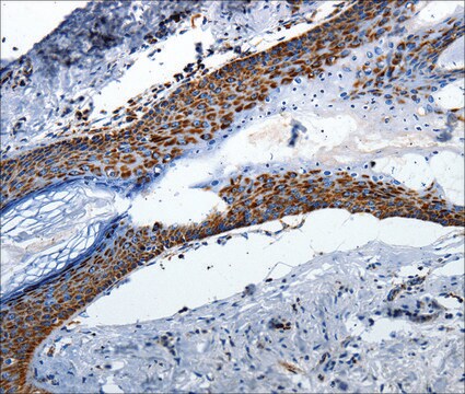
Immunohistochemistry Analysis: 1:500 dilution from a previous lot detected Slo1 in rat cerebellum tissue.

Immunocytochemistry Analysis: A previous lot was used by an independent laboratory in IC. (Misonou, H., et al. (2006). J Comp Neurol. 496(3):289–302.)
Research Category
Neuroscience
Research Sub Category
Signaling Neuroscience

Quality

Evaluated by Western Blot in rat brain membrane tissue lysate.

Western Blot Analysis: 1 µg/mL of this antibody detected Slo1 on 10 µg of rat brain membrane tissue lysate.

Target description

~100 kDa observed

Physical form

Format: Purified
Protein G
Purified mouse monoclonal IgG2aκ in buffer containing 0.1 M Tris-Glycine (pH 7.4), 150 mM NaCl with 0.05% sodium azide.

Storage and Stability

Stable for 1 year at 2-8°C from date of receipt.

Analysis Note

Control
Rat brain membrane tissue lysate

Other Notes

Concentration: Please refer to the Certificate of Analysis for the lot-specific concentration.

Disclaimer

Unless otherwise stated in our catalog or other company documentation accompanying the product(s), our products are intended for research use only and are not to be used for any other purpose, which includes but is not limited to, unauthorized commercial uses, in vitro diagnostic uses, ex vivo or in vivo therapeutic uses or any type of consumption or application to humans or animals.

Not finding the right product?  

Try our Product Selector Tool.

Storage Class Code

12 - Non Combustible Liquids

WGK

WGK 1

Flash Point(F)

Not applicable

Flash Point(C)

Not applicable


Certificates of Analysis (COA)

Search for Certificates of Analysis (COA) by entering the products Lot/Batch Number. Lot and Batch Numbers can be found on a product’s label following the words ‘Lot’ or ‘Batch’.

Already Own This Product?

Find documentation for the products that you have recently purchased in the Document Library.

Visit the Document Library

Boxing Li et al.
Nature neuroscience, 17(8), 1055-1063 (2014-06-24)
Ion channels are essential for the regulation of neuronal functions. The significance of plasma membrane, mitochondrial, endoplasmic reticulum and lysosomal ion channels in the regulation of Ca(2+) is well established. In contrast, surprisingly little is known about the function of
Md Azadul Kabir Sarker et al.
Endocrinology, 160(1), 235-248 (2018-11-27)
Class II phosphoinositide 3-kinases (PI3Ks), PI3K-C2α and PI3K-C2β, are highly homologous and distinct from class I and class III PI3Ks in catalytic products and domain structures. In contrast to class I and class III PI3Ks, physiological roles of PI3K-C2α and

Our team of scientists has experience in all areas of research including Life Science, Material Science, Chemical Synthesis, Chromatography, Analytical and many others.

Contact Technical Service