AB5330
Anti-Syntaxin 4 Antibody
Chemicon®, from rabbit
Sign Into View Organizational & Contract Pricing
All Photos(1)
About This Item
UNSPSC Code:
12352203
eCl@ss:
32160702
NACRES:
NA.41
Recommended Products
biological source
rabbit
Quality Level
antibody form
affinity purified immunoglobulin
antibody product type
primary antibodies
clone
polyclonal
purified by
affinity chromatography
species reactivity
rat
manufacturer/tradename
Chemicon®
technique(s)
western blot: suitable
NCBI accession no.
UniProt accession no.
shipped in
wet ice
target post-translational modification
unmodified
Gene Information
human ... STX4(6810)
Specificity
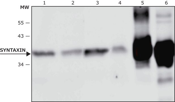
Recognizes Syntaxin 4.
Immunogen
Highly purified corresponding to residues 2-23 of rat or mouse Syntaxin 4 (Accession Q08850).
Application
Research Category
Neuroscience
Neuroscience
Research Sub Category
Synapse & Synaptic Biology
Synapse & Synaptic Biology
This Anti-Syntaxin 4 Antibody is validated for use in WB for the detection of Syntaxin 4.
Western blotting: 0.5-1.0 μg/mL (1:200-1:500) Dilutions should be made using a carrier protein such as BSA (1-3%).
Optimal working dilutions must be determined by the end user.
Optimal working dilutions must be determined by the end user.
Physical form
Affinity purified immunoglobulin. Lyophilized from PBS, pH 7.4, containing 1% BSA and 0.025% sodium azide. Reconstitute with 200 μL of sterile distilled water. Centrifuge antibody preparation before use (10,000 x g for 5 min).
Storage and Stability
Maintain lyophilized material at -20°C for up to 6 months. After reconstitution maintain at -20°C in undiluted aliquots for up to 6 months. Avoid repeated freeze/thaw cycles.
Analysis Note
Control
CONTROL ANTIGEN: Included free of charge with the antibody is 40 μg of control antigen (lyophilized powder). Reconstitute with 100 μL of sterile distilled water. For negative control, preincubate 1 μg of purified peptide with 1 μg of antibody for one hour at room temperature. Optimal concentrations must be determined by the end user.
CONTROL ANTIGEN: Included free of charge with the antibody is 40 μg of control antigen (lyophilized powder). Reconstitute with 100 μL of sterile distilled water. For negative control, preincubate 1 μg of purified peptide with 1 μg of antibody for one hour at room temperature. Optimal concentrations must be determined by the end user.
Other Notes
Concentration: Please refer to the Certificate of Analysis for the lot-specific concentration.
Legal Information
CHEMICON is a registered trademark of Merck KGaA, Darmstadt, Germany
Disclaimer
Unless otherwise stated in our catalog or other company documentation accompanying the product(s), our products are intended for research use only and are not to be used for any other purpose, which includes but is not limited to, unauthorized commercial uses, in vitro diagnostic uses, ex vivo or in vivo therapeutic uses or any type of consumption or application to humans or animals.
Not finding the right product?
Try our Product Selector Tool.
Hazard Statements
Precautionary Statements
Hazard Classifications
Aquatic Chronic 3
Storage Class Code
11 - Combustible Solids
WGK
WGK 3
Certificates of Analysis (COA)
Search for Certificates of Analysis (COA) by entering the products Lot/Batch Number. Lot and Batch Numbers can be found on a product’s label following the words ‘Lot’ or ‘Batch’.
Already Own This Product?
Find documentation for the products that you have recently purchased in the Document Library.
Arlene A Hirano et al.
Visual neuroscience, 24(4), 489-502 (2007-07-21)
Horizontal cells mediate inhibitory feed-forward and feedback communication in the outer retina; however, mechanisms that underlie transmitter release from mammalian horizontal cells are poorly understood. Toward determining whether the molecular machinery for exocytosis is present in horizontal cells, we investigated
Eunjin Oh et al.
Diabetes, 70(12), 2837-2849 (2021-09-25)
Syntaxin 4 (STX4), a plasma membrane-localized SNARE protein, regulates human islet β-cell insulin secretion and preservation of β-cell mass. We found that human type 1 diabetes (T1D) and NOD mouse islets show reduced β-cell STX4 expression, consistent with decreased STX4
Christian Puller et al.
PloS one, 9(2), e88963-e88963 (2014-03-04)
The functional roles and synaptic features of horizontal cells in the mammalian retina are still controversial. Evidence exists for feedback signaling from horizontal cells to cones and feed-forward signaling from horizontal cells to bipolar cells, but the details of the
M Joseph Phillips et al.
The Journal of comparative neurology, 518(11), 2071-2089 (2010-04-16)
The Pde6b(rd10) (rd10) mouse has a moderate rate of photoreceptor degeneration and serves as a valuable model for human autosomal recessive retinitis pigmentosa (RP). We evaluated the progression of neuronal remodeling of second- and third-order retinal cells and their synaptic
Our team of scientists has experience in all areas of research including Life Science, Material Science, Chemical Synthesis, Chromatography, Analytical and many others.
Contact Technical Service