Skip to Content
Merck
All Photos(3)

Key Documents

SAB4200643

Sigma-Aldrich

Monoclonal Anti-PLEKHA7 antibody produced in mouse

clone PLK7, purified from hybridoma cell culture

Sign Into View Organizational & Contract Pricing


About This Item

UNSPSC Code:
12352203
NACRES:
NA.41
clone:
PLK7, monoclonal
application:
FACS
ICC
species reactivity:
human
technique(s):
flow cytometry: 2-4 μg/test using NCI-H1299 cells
immunoblotting: suitable
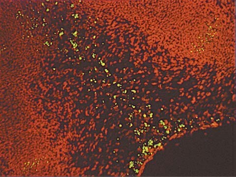
immunocytochemistry: 0.25-0.5 μg/mL using human CaCo2 cells
citations:
3

biological source

mouse

Quality Level

antibody form

purified immunoglobulin

antibody product type

primary antibodies

clone

PLK7, monoclonal

form

buffered aqueous solution

species reactivity

human

concentration

~1 mg/mL

technique(s)

flow cytometry: 2-4 μg/test using NCI-H1299 cells
immunoblotting: suitable
immunocytochemistry: 0.25-0.5 μg/mL using human CaCo2 cells

isotype

IgG2a

UniProt accession no.

shipped in

dry ice

storage temp.

−20°C

target post-translational modification

unmodified

Gene Information

Related Categories

General description

Monoclonal Anti-PLEKHA7 (mouse IgG2a isotype) is derived from the hybridoma PLK7 produced by the fusion of mouse myeloma cells and splenocytes from BALB/c mice immunized with a synthetic peptide corresponding to a sequence at the N-terminal region of human PLEKHA7, conjugated to KLH. PLEKHA7 contains WW (rsp5-domain), PH (pleckstrin homology), and CC (coiled-coil) domains.
PLEKHA7 (Pleckstrin homology domain containing, family A member 7) is an adherens junction protein localized at the adherens junctions in colonic epithelial cells.

Immunogen

synthetic peptide corresponding to a sequence at the N-terminal region of human PLEKHA7

Application

Monoclonal Anti-PLEKHA7 antibody has been used in
  • immunoblotting
  • immunocytochemistry
  • flow cytometry

Biochem/physiol Actions

Overexpression of PLEKHA7 was reported to be common in invasive lobular carcinomas (ILCs) and hence PLEKHA7 could serve as a molecular marker to differentiate ILCs from invasive ductal carcinomas (IDCs).
PLEKHA7 (Pleckstrin homology domain containing, family A member 7) functions as a cytoplasmic component of the epithelial adherens junction belt, with a subcellular localization and tissue distribution and connects E-cadherin-p120 ctn complex to the microtubule cytoskeleton. It has been reported that PLEKHA7 acts as zonula adherens (ZA) component, identifies Nezha, which further binds to microtubules (MTs) of the cytoskeleton, and forms a protein complex during cytoskeleton assembly.

Physical form

Solution in 0.01 M phosphate buffered saline, pH 7.4, containing 15 mM sodium azide.

Disclaimer

Unless otherwise stated in our catalog or other company documentation accompanying the product(s), our products are intended for research use only and are not to be used for any other purpose, which includes but is not limited to, unauthorized commercial uses, in vitro diagnostic uses, ex vivo or in vivo therapeutic uses or any type of consumption or application to humans or animals.

Not finding the right product?  

Try our Product Selector Tool.

Storage Class Code

10 - Combustible liquids

WGK

WGK 1

Flash Point(F)

Not applicable

Flash Point(C)

Not applicable


Choose from one of the most recent versions:

Certificates of Analysis (COA)

Lot/Batch Number

Don't see the Right Version?

If you require a particular version, you can look up a specific certificate by the Lot or Batch number.

Already Own This Product?

Find documentation for the products that you have recently purchased in the Document Library.

Visit the Document Library

Anna Akhmanova et al.
Cell, 135(5), 791-793 (2008-12-02)
The zonula adherens (ZA) is a specialized cadherin-based structure found at the contacts between epithelial cells. Meng et al. (2008) now identify a protein complex containing the microtubule minus-end-binding protein Nezha, which provides a critical link between microtubules and cadherins
Souichi Kurita et al.
The Journal of biological chemistry, 288(41), 29356-29368 (2013-08-31)
Adherens junction (AJ) is a specialized cell-cell junction structure that plays a role in mechanically connecting adjacent cells to resist strong contractile forces and to maintain tissue structure, particularly in the epithelium. AJ is mainly comprised of cell adhesion molecules
Bàrbara Castellana et al.
Human pathology, 43(11), 1902-1909 (2012-05-01)
Molecular differentiation between invasive lobular carcinomas (ILCs) and invasive ductal carcinomas (IDCs) of the breast has not been well defined. We investigated gene expression differences between ILCs and IDCs and their correlation with variations in invasiveness and tumor growth. Total
Pamela Pulimeno et al.
PloS one, 5(8), e12207-e12207 (2010-09-03)
The pleckstrin-homology-domain-containing protein PLEKHA7 was recently identified as a protein linking the E-cadherin-p120 ctn complex to the microtubule cytoskeleton. Here we characterize the expression, tissue distribution and subcellular localization of PLEKHA7 by immunoblotting, immunofluorescence microscopy, immunoelectron microscopy, and northern blotting
Wenxiang Meng et al.
Cell, 135(5), 948-959 (2008-12-02)
Epithelial cells contain noncentrosomal microtubules (MTs), whose minus ends are oriented apically. In contrast with the well-known interactions of the minus ends with the centrosome, little is known about the termination site of the noncentrosomal minus ends. Here we show

Our team of scientists has experience in all areas of research including Life Science, Material Science, Chemical Synthesis, Chromatography, Analytical and many others.

Contact Technical Service