Skip to Content
Merck
All Photos(1)

Key Documents

N0913

Sigma-Aldrich

Anti-Neurocan antibody, Mouse monoclonal

clone 650.24, purified from hybridoma cell culture

Synonym(s):

Anti-CSPG3, Anti-Chondroitin Sulfate Proteogycan 3

Sign Into View Organizational & Contract Pricing


About This Item

MDL number:
UNSPSC Code:
12352203
NACRES:
NA.41

biological source

mouse

Quality Level

conjugate

unconjugated

antibody form

purified from hybridoma cell culture

antibody product type

primary antibodies

clone

650.24, monoclonal

form

buffered aqueous solution

mol wt

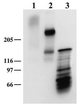
antigen 260 kDa (additional band at 160 kDa)

species reactivity

rat

packaging

antibody small pack of 25 μL

concentration

~1 mg/mL

technique(s)

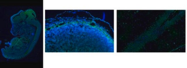
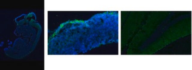
immunocytochemistry: suitable
immunohistochemistry (frozen sections): suitable
indirect ELISA: suitable
western blot: 0.1-0.2 μg/mL using neonatal rat brain treated with chondroitinase.

isotype

IgG1

UniProt accession no.

shipped in

dry ice

storage temp.

−20°C

target post-translational modification

unmodified

Gene Information

rat ... Ncan(58982)

Related Categories

General description

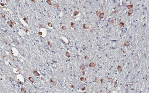
Monoclonal Anti-Neurocan (mouse IgG1 isotype) is derived from the hybridoma 650.24 produced by the fusion of mouse myeloma cells (P3X cells) and splenocytes from BALB/c mice immunized with embryonic rat brain proteoglycans. Neurocan is a chondroitin sulfate proteoglycan (known also as CSPG3). It accounts for 20−30% of chondroitin sulfate proteoglycans in early postnatal rat brain. The mouse neurocan cDNA encodes for a 1,257-amino acid protein with a 22-amino acid signal peptide followed by an immunoglobulin-like domain and repeating motifs characteristic of the hyaluronic acid-binding region of aggregating proteoglycans. Neurocan is found in large amounts in glial scar, a primarily astrocytic structure formed after an injury in the central nervous system (CNS).
Neurocan is a proteoglycan which is expressed in the nervous tissue.

Immunogen

embryonic rat brain proteoglycans.

Application

Anti-Neurocan antibody, Mouse monoclonal has been used in:
  • western blotting
  • immunofluorescence
  • enzyme-linked immunosorbent assay (ELISA)
  • immunocytochemistry
  • immunohistochemistry

Biochem/physiol Actions

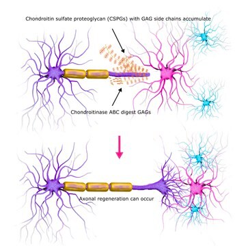
Neurocan can inhibit the growth of axons. In presence of central nervous system injuries, it also takes part in tissue repair and neural network reconstruction.
Neurocan participates in the modulation of cell adhesion and migration. It exerts a negative effect on growing cerebellar axons.

Target description

Neurocan is a chondroitin sulfate proteoglycan (knownalso as CSPG3) thought to be involved in the modulation of cell adhesion and migration. It accounts for 20-30% of chondroitin sulfate proteoglycans in early postnatal rat brain.

Physical form

Solution in 0.01 M phosphate buffered saline, pH 7.4, containing 15 mM sodium azide.

Disclaimer

Unless otherwise stated in our catalog or other company documentation accompanying the product(s), our products are intended for research use only and are not to be used for any other purpose, which includes but is not limited to, unauthorized commercial uses, in vitro diagnostic uses, ex vivo or in vivo therapeutic uses or any type of consumption or application to humans or animals.

Not finding the right product?  

Try our Product Selector Tool.

Storage Class Code

10 - Combustible liquids

WGK

WGK 3

Flash Point(F)

Not applicable

Flash Point(C)

Not applicable


Certificates of Analysis (COA)

Search for Certificates of Analysis (COA) by entering the products Lot/Batch Number. Lot and Batch Numbers can be found on a product’s label following the words ‘Lot’ or ‘Batch’.

Already Own This Product?

Find documentation for the products that you have recently purchased in the Document Library.

Visit the Document Library

AMPA Receptor Antagonist NBQX Decreased Seizures by Normalization of Perineuronal Nets
Chen W, et al.
PLoS ONE, 11(11), e0166672-e0166672 (2016)
Neurocan: a brain chondroitin sulfate proteoglycan
Rauch U, et al.
Cellular and Molecular Life Sciences, 58(12-13), 1842-1856 (2001)
Changes in neurocan expression in the distal spinal cord stump following complete cord transection: a comparison between infant and adult rats.
Qi ML
Neuroscience Research, 45(2), 181-188 (2003)
Characterization of the human neurocan gene, CSPG3
Prange CK, et al.
Gene, 221(2), 199-205 (1998)
Li Hong Shen et al.
Glia, 56(16), 1747-1754 (2008-07-12)
The glial scar, a primarily astrocytic structure bordering the infarct tissue inhibits axonal regeneration after stroke. Neurocan, an axonal extension inhibitory molecule, is up-regulated in the scar region after stroke. Bone marrow stromal cells (BMSCs) reduce the thickness of glial

Our team of scientists has experience in all areas of research including Life Science, Material Science, Chemical Synthesis, Chromatography, Analytical and many others.

Contact Technical Service