F1773
Anti-CD4−FITC antibody, Mouse monoclonal
clone Q4120, purified from hybridoma cell culture
Synonym(s):
Monoclonal Anti-CD4
Sign Into View Organizational & Contract Pricing
All Photos(1)
About This Item
UNSPSC Code:
12352203
NACRES:
NA.44
Recommended Products
biological source
mouse
Quality Level
conjugate
FITC conjugate
antibody form
purified immunoglobulin
antibody product type
primary antibodies
clone
Q4120, monoclonal
form
buffered aqueous solution
mol wt
antigen 59 kDa
species reactivity
human
technique(s)
flow cytometry: 10 μL using 1 × 10⁶ cells
isotype
IgG1
UniProt accession no.
shipped in
wet ice
storage temp.
2-8°C
target post-translational modification
unmodified
Gene Information
human ... CD4(920)
Looking for similar products? Visit Product Comparison Guide
General description
Anti-CD4-FITC antibody, mouse monoclonal (mouse IgG1 isotype) is derived from the hybridoma produced by the fusion of mouse myeloma cell line NS-1 and splenocytes from Balb/c mice immunized with cluster of differentiation 4 (CD4)-transfected mouse T cell hybridoma, 3DT, followed by CD4+ human T cell CEM cells (leukemic lymphoblasts).
CD4, a glycoprotein, is predominantly expressed on the cell membrane of mature, thymus-derived (T) lymphocyte and also to some extent in the monocyte/macrophage lineage. It has a transmembrane domain, cytoplasmic region and an extracellular region of four tandem domain similar like members of Ig superfamily.
Specificity
Recognizes the CD4. The epitope recognized by the Q4120 clone is located on 1-183 aa and is sensitive to formalin fixation and paraffin embedding. 5th Workshop: code no. CD04.11
Immunogen
CD4-transfected mouse T-cell hybridoma, 3DT, followed by CD4+ human T-cell CEM cells.
Application
FITC Conjugated Monoclonal Anti-Human CD4 has been used for:
- identification, quantification and monitoring of helper/inducer T cells in peripheral blood, biological fluids, lymphoid organs, and other tissues
- analysis of T cell mediated cytotoxicity
- characterization of subtypes of T cell leukemias and lymphomas
- studies of T cells in health and disease
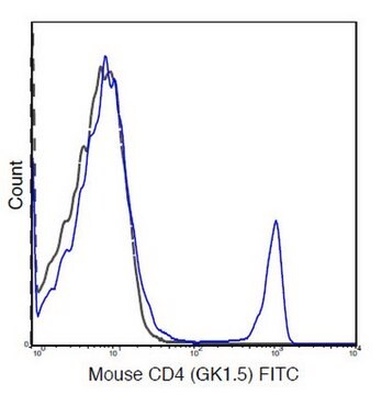
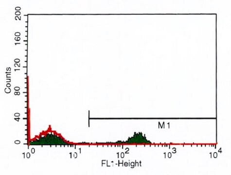
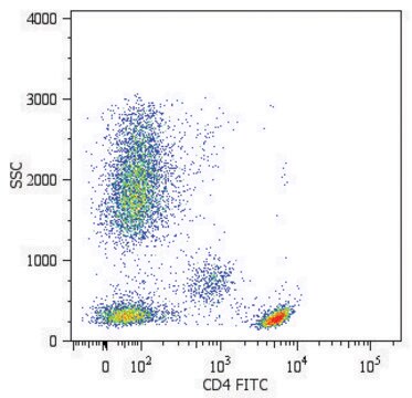
- flow cytometry
Biochem/physiol Actions
CD4+ T cells acts as a helper/inducer, in providing an activating signal to B cells. It induces T lymphocytes bearing the reciprocal CD8 marker to become cytotoxic/suppressor cells. It is also majorly associated with non-polymorphic region of MHC (major histocompatibility complex) antigen in interacting with targets bearing MHC class II molecules. Thus, it is called as an adhesion molecule. It also behaves as receptor for the GP 120 envelope glycoprotein of human immunodeficiency virus. The CD4 molecules is involved in the adhesion of T lymphocytes to target cells, thymic development, transmission of intracellular signals during T cell activation, and binding to polyclonal immunoglobulins.
Immunoregulatory T cell subset abnormalities in autoimmunity disorders, immunodeficiency diseases, graft versus host disease and following immunosuppressive therapy are often manifested as a change in CD4+/CD8+ ratio in peripheral blood T cells.
Target description
CD4 is a single chain transmembraneous glycoprotein from the immunoglobulin superfamily. It is expressed on the helper/inducer T subset, on most medullary thymocytes, on microglial, dendritic and on some malignancies of T cell origin. The antigen binds to MHC class II molecules and is associated with p56lck protein tyrosine kinase.
Physical form
Solution in 0.01 M phosphate buffered saline, pH 7.4, containing 1% bovine serum albumin and 15 mM sodium azide.
Preparation Note
Prepared by conjugation to fluorescein isothiocyanate isomer I (FITC). This green dye is efficiently excited at 495 nm and emits at 525 nm.
Disclaimer
Unless otherwise stated in our catalog or other company documentation accompanying the product(s), our products are intended for research use only and are not to be used for any other purpose, which includes but is not limited to, unauthorized commercial uses, in vitro diagnostic uses, ex vivo or in vivo therapeutic uses or any type of consumption or application to humans or animals.
Not finding the right product?
Try our Product Selector Tool.
Storage Class Code
10 - Combustible liquids
WGK
nwg
Flash Point(F)
Not applicable
Flash Point(C)
Not applicable
Personal Protective Equipment
dust mask type N95 (US), Eyeshields, Gloves
Choose from one of the most recent versions:
Already Own This Product?
Find documentation for the products that you have recently purchased in the Document Library.
The production of armed effector T cells
Janeway, et al.
Immunobiology (2001)
Qi-Zhi Zhang et al.
Molecular medicine reports, 22(5), 3629-3634 (2020-10-02)
Phosphoinositide 3-kinase catalytic subunit δ isoform (P110δ) is mainly expressed in white blood cells. It is involved in T and B lymphocyte differentiation, maturation and the neutrophil chemotaxis process. Apolipoprotein E (ApoE) is an arginine‑rich alkaline protein, which is present
Variation of blood T lymphocyte subgroups in patients with non-small cell lung cancer
Wang WJ, et al.
Asian Pacific Journal of Cancer Prevention, 14(8), 4671-4673 (2013)
Ravi Tandon et al.
AIDS research and human retroviruses, 30(7), 654-664 (2014-05-03)
Galectin-9 (Gal-9) is a β-galactosidase-binding lectin that promotes apoptosis, tissue inflammation, and T cell immune exhaustion, and alters HIV infection in part through engagement with the T cell immunoglobulin mucin domain-3 (Tim-3) receptor and protein disulfide isomerases (PDI). Gal-9 was
The CD4 antigen: physiological ligand and HIV receptor.
Q J Sattentau et al.
Cell, 52(5), 631-633 (1988-03-11)
Our team of scientists has experience in all areas of research including Life Science, Material Science, Chemical Synthesis, Chromatography, Analytical and many others.
Contact Technical Service