All Photos(1)
About This Item
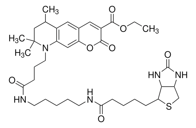
Empirical Formula (Hill Notation):
C46H57ClN6O10S
Molecular Weight:
921.50
MDL number:
UNSPSC Code:
12352108
NACRES:
NA.32
Recommended Products
Assay
90% (HPLC)
Quality Level
manufacturer/tradename
ATTO-TEC GmbH
fluorescence
λex 565 nm; λem 590 nm in 0.1 M phosphate pH 7.0
λ
in ethanol (with 0.1% trifluoroacetic acid)
storage temp.
−20°C
Storage Class Code
11 - Combustible Solids
WGK
WGK 3
Flash Point(F)
Not applicable
Flash Point(C)
Not applicable
Personal Protective Equipment
dust mask type N95 (US), Eyeshields, Gloves
Choose from one of the most recent versions:
Already Own This Product?
Find documentation for the products that you have recently purchased in the Document Library.
Customers Also Viewed
Suman Lata et al.
Journal of the American Chemical Society, 128(7), 2365-2372 (2006-02-16)
Labeling of proteins with fluorescent dyes offers powerful means for monitoring protein interactions in vitro and in live cells. Only a few techniques for noncovalent fluorescence labeling with well-defined localization of the attached dye are currently available. Here, we present
Łukasz Krzemiński et al.
Journal of the American Chemical Society, 133(38), 15085-15093 (2011-08-26)
A combined fluorescence and electrochemical method is described that is used to simultaneously monitor the type-1 copper oxidation state and the nitrite turnover rate of a nitrite reductase (NiR) from Alcaligenes faecalis S-6. The catalytic activity of NiR is measured
Marie-Luise Humpert et al.
Proteomics, 12(12), 1938-1948 (2012-05-25)
PTMs of extracellular domains of membrane proteins can influence antibody binding and give rise to ambivalent results. Best proof of protein expression is the use of complementary methods to provide unequivocal evidence. CXCR7, a member of the atypical chemokine receptor
Takao Nakata et al.
The Journal of cell biology, 194(2), 245-255 (2011-07-20)
Polarized transport in neurons is fundamental for the formation of neuronal circuitry. A motor domain-containing truncated KIF5 (a kinesin-1) recognizes axonal microtubules, which are enriched in EB1 binding sites, and selectively accumulates at the tips of axons. However, it remains
Ashwanth C Francis et al.
Cell host & microbe, 23(4), 536-548 (2018-04-13)
The HIV-1 core consists of capsid proteins (CA) surrounding viral genomic RNA. After virus-cell fusion, the core enters the cytoplasm and the capsid shell is lost through uncoating. CA loss precedes nuclear import and HIV integration into the host genome
Our team of scientists has experience in all areas of research including Life Science, Material Science, Chemical Synthesis, Chromatography, Analytical and many others.
Contact Technical Service


