
MABS827
Anti-ERK1/2 Antibody, clone 16A6.1
clone 16A6.1, from mouse
Synonym(s):
EC: 2.7.11.24, MAP kinase 3, MAPK3, ERT2, ERK-1, Insulin-stimulated MAP2 kinase, MAP kinase isoform p44, p44-MAPK, MNK1, Microtubule-associated protein 2 kinase, p44-ERK1
About This Item
Recommended Products
biological source
mouse
Quality Level
antibody form
purified antibody
antibody product type
primary antibodies
clone
16A6.1, monoclonal
species reactivity
rat, human, mouse
technique(s)
immunohistochemistry: suitable (paraffin)
western blot: suitable
isotype
IgG2bκ
NCBI accession no.
UniProt accession no.
shipped in
ambient
target post-translational modification
unmodified
Gene Information
human ... MAPK3(5595)
mouse ... Mapk3(26417)
rat ... Mapk3(50689)
General description
Specificity
Immunogen
Application
Signaling
Quality
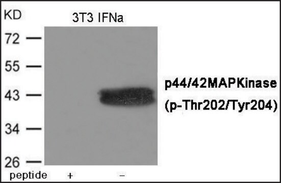
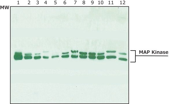
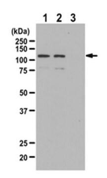
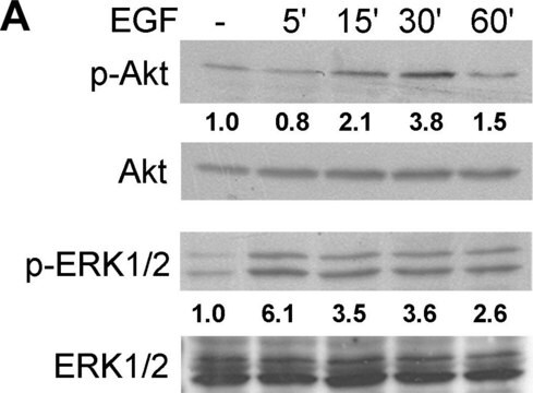
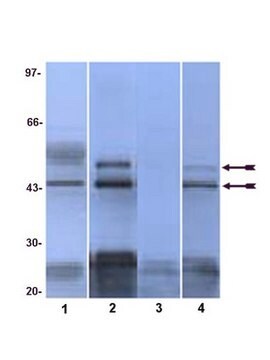
Western Blotting Analysis: 0.5 µg/mL of this antibody detected ERK1/2 in 10 µg of RAW264.7 cell lysate.
Target description
Linkage
Physical form
Storage and Stability
Other Notes
Disclaimer
Not finding the right product?
Try our Product Selector Tool.
recommended
Storage Class Code
12 - Non Combustible Liquids
WGK
WGK 1
Certificates of Analysis (COA)
Search for Certificates of Analysis (COA) by entering the products Lot/Batch Number. Lot and Batch Numbers can be found on a product’s label following the words ‘Lot’ or ‘Batch’.
Already Own This Product?
Find documentation for the products that you have recently purchased in the Document Library.
Our team of scientists has experience in all areas of research including Life Science, Material Science, Chemical Synthesis, Chromatography, Analytical and many others.
Contact Technical Service