Skip to Content
Merck
All Photos(1)

Key Documents

MABF2070

Sigma-Aldrich

Anti-Protein-tyrosine phosphatase eta (CD148) Antibody, clone 8A-1

clone 8A-1, from hamster(Syrian)

Synonym(s):

Receptor-type tyrosine-protein phosphatase eta, EC: 3.1.3.48, Protein-tyrosine phosphatase eta, R-PTP-eta, HPTP beta-like tyrosine phosphatase, Protein-tyrosine phosphatase receptor type J, R-PTP-J, Susceptibility to colon cancer 1, CD148

Sign Into View Organizational & Contract Pricing


About This Item

UNSPSC Code:
12352203
eCl@ss:
32160702
NACRES:
NA.41

biological source

hamster (Syrian)

Quality Level

antibody form

purified antibody

antibody product type

primary antibodies

clone

8A-1, monoclonal

species reactivity

mouse

packaging

antibody small pack of 25 μg

technique(s)

flow cytometry: suitable
immunohistochemistry: suitable (paraffin)
western blot: suitable

NCBI accession no.

UniProt accession no.

shipped in

ambient

target post-translational modification

unmodified

Gene Information

mouse ... Ptprj(19271)

Related Categories

General description

Receptor-type tyrosine-protein phosphatase eta (UniProt: Q64455, also known as EC: 3.1.3.48, Protein-tyrosine phosphatase eta, R-PTP-eta, HPTP beta-like tyrosine phosphatase Protein-tyrosine phosphatase receptor type J, R-PTP-J, Susceptibility to colon cancer 1, CD148) is encoded by the Ptprj (also known as Byp, Scc1) gene (Gene ID: 19271) in murine species. R-PTP-eta is a monomeric, single pass type I membrane protein that is expressed at high levels in brain, kidney, spleen, and intestine; Lower expression levels are seen in liver, lung, thymus, and heart. It is also expressed at a high level in the myeloid cell line FDC-P2, and at a lower level in the pre-B lymphoid cell line WEHI-231. Fibroblast cell line NIH3T3 and the erythroid cell line F5-5 lack its expression. It is synthesized with a signal peptide (aa 1-28) that is subsequently cleaved off. The mature form has an extracellular domain (aa 29-876), a helical region (aa 877-897), a cytoplasmic domain (aa 898-1238). R-PTP-eta either dephosphorylates or contributes to the dephosphorylation of CTNND1, FLT3, PDGFRB, MET, RET, KDR, LYN, SRC, MAPK1, MAPK3, EGFR, TJP1, OCLN, PIK3R1 and PIK3R2 and plays a role in cell adhesion, migration, proliferation and differentiation. It is also involved in vascular development and ma participate in the mechanism of contact inhibition of cell growth. It has a positive effect on cell-matrix adhesion and on platelet activation and thrombosis. It can also serve as a negative regulator of cell proliferation and has a negative effect on PDGF-stimulated cell migration via dephosphorylation of PDGFR. R-PTP-eta is also involved in T cell receptor (TCR) signaling where upon TCR activation, it is up-regulated and excluded from the immunological synapses, while upon T cell-antigen presenting cells (APC) disengagement, it is no longer excluded and can dephosphorylate PLCG1 and LAT to down-regulate prolongation of signaling.

Specificity

Clone 8A-1 immunostains Protein-tyrosine phosphatase eta in murine spleen tissue secctions.

Immunogen

293T cells expressing murine CD148.

Application

Anti-Protein-tyrosine phosphatase eta (CD148), clone 8A-1, Cat. No. MABF2070, is a Syrian hamster monoclonal antibody that detects Receptor-type tyrosine-protein phosphatase eta (CD148) and is tested for use in Flow Cytometry, Immunohistochemistry (Paraffin), and Western Blotting.
Research Category
Inflammation & Immunology
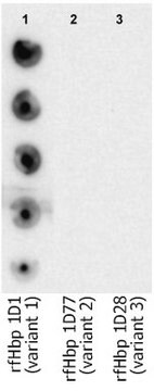
Western Blotting Analysis: A representative lot detected Protein-tyrosine phosphatase eta (CD148) in Western Blotting applications (Stepanek, O., et. al. (2011). J Biol Chem. 286(25):22101-12).

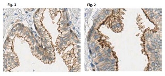
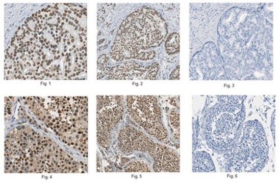
Immunohistochemistry Analysis: A representative lot detected Protein-tyrosine phosphatase eta (CD148) in Immunohistochemistry applications (Katsumoto, T.R., et. al. (2013). J Clin Invest. 123(5):2037-48).

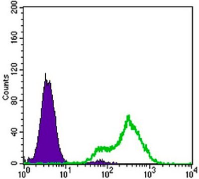
Flow Cytometry Analysis: A representative lot detected Protein-tyrosine phosphatase eta (CD148) in Flow Cytometry applications (Stepanek, O., et. al. (2011). J Biol Chem. 286(25):22101-12; Lin, J., et. al. (2004). J Immunol. 173(4):2324-30).

Quality

Evaluated by Immunohistochemistry in mouse spleen tissue.

Immunohistochemistry Analysis: A 1:250 dilution of this antibody immunostained Protein-tyrosine phosphatase eta (CD148) in mouse spleen tissue sections.

Target description

136.77 kDa calculated.

Physical form

Format: Purified
Protein G purified
Purified Syrian hamster monoclonal antibody IgG2-IgG3 in buffer containing 0.1 M Tris-Glycine (pH 7.4), 150 mM NaCl with 0.05% sodium azide.

Storage and Stability

Stable for 1 year at 2-8°C from date of receipt.

Other Notes

Concentration: Please refer to lot specific datasheet.

Disclaimer

Unless otherwise stated in our catalog or other company documentation accompanying the product(s), our products are intended for research use only and are not to be used for any other purpose, which includes but is not limited to, unauthorized commercial uses, in vitro diagnostic uses, ex vivo or in vivo therapeutic uses or any type of consumption or application to humans or animals.

Not finding the right product?  

Try our Product Selector Tool.


Certificates of Analysis (COA)

Search for Certificates of Analysis (COA) by entering the products Lot/Batch Number. Lot and Batch Numbers can be found on a product’s label following the words ‘Lot’ or ‘Batch’.

Already Own This Product?

Find documentation for the products that you have recently purchased in the Document Library.

Visit the Document Library

Our team of scientists has experience in all areas of research including Life Science, Material Science, Chemical Synthesis, Chromatography, Analytical and many others.

Contact Technical Service