Skip to Content
Merck
All Photos(4)

Key Documents

MABE1122

Sigma-Aldrich

Anti-HOIP (RNF31) Antibody, clone 1CB2

clone 1CB2, from mouse

Synonym(s):

E3 ubiquitin-protein ligase RNF31, HOIL-1-interacting protein, HOIP, RING finger protein 31, Zinc in-between-RING-finger ubiquitin-associated domain protein

Sign Into View Organizational & Contract Pricing


About This Item

UNSPSC Code:
12352203
eCl@ss:
32160702
NACRES:
NA.41
clone:
1CB2, monoclonal
application:
IHC
WB
species reactivity:
human
technique(s):
immunohistochemistry: suitable (paraffin)
western blot: suitable
citations:

biological source

mouse

Quality Level

antibody form

purified immunoglobulin

antibody product type

primary antibodies

clone

1CB2, monoclonal

species reactivity

human

should not react with

mouse

technique(s)

immunohistochemistry: suitable (paraffin)
western blot: suitable

isotype

IgG1κ

NCBI accession no.

UniProt accession no.

shipped in

ambient

target post-translational modification

unmodified

Gene Information

human ... RNF31(55072)

General description

E3 ubiquitin-protein ligase RNF31 (UniProt Q96EP0; also known as HOIL-1-interacting protein, HOIP, RING finger protein 31, Zinc in-between-RING-finger ubiquitin-associated domain protein) is encoded by the RNF31 (also known as ZIBRA) gene (Gene ID 55072) in human. HOIP is a RING-between-RING (RBR) family E3 ubiquitin ligase and the only enzyme known to catalyze linear-ubiquitin-chain conjugation. HOIP functions as the primary E3 Ub ligase of the immune signaling regulatory complex LUBAC (linear-ubiquitin-chain assembly complex) composed of heme-oxidized IRP2 ubiquitin ligase-1 (HOIL-1L), HOIP, and SHANK-associated RH domain interactor (Sharpin). RBR ligases often exist in autoinhibited conformations and require protein-protein interactions or posttranslational modifications to induce a conformational change for activation. Disruption of HOIP and HOIL-1L interaction in activated B cell-like (ABC) subtype of diffuse large B-cell lymphoma (DLBCL) induces DLBCL death as a result of decreased NF-κB activation due to HOIP inactivation. On the other hand, HOIP K1056R mutant is resistant to inactivation by TLR4 stimulation-induced HOIP K1056 ubiquitination. Human HOIP is a 1072-a.a. protein that contains a polyUb-binding region (a.a. 1-485), a ubiquitin-associated or UBA domain (a.a. 564-615) that mediates HOIL-1L/RBCK1 interactioin, and a C-terminal linear-ubiquitin-chain-determining domain or LDD domain (a.a. 910-1072). In addition, HOIP conatins three RanBP2-type Zinc fingers (a.a. 299-329, 350-379, 409-438), two RING-type Zinc fingers (a.a. 699-747 and 860-909), and one IBR-type Zinc fingers (a.a. 779-841).

Specificity

Clone 1CB2 detected a ~120 kDa target band only in parental, but not HOIP-knock, Jurkat cells. Clone 1CB2 targets an epitope within the C-terminal LDD domain present in all three human HOIP (RNF31) spliced isoforms reported by UniProt (Q96EP0).

Immunogen

GST-tagged recombinant human HOIP (RNF31) LDD domain.

Application

Detect HOIP using this mouse monoclonal Anti-HOIP (RNF31), clone 1CB2 Antibody, Cat. No. MABE1122, validated for use in Immunohistochemistry (Paraffin) and Western Blotting.
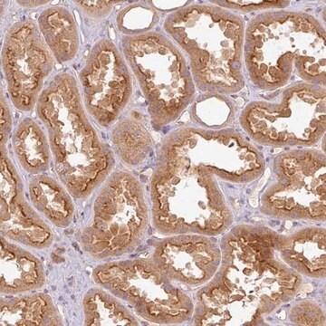
Immunohistochemistry Analysis: A 1:10 dilution from a representative lot detected HOIP (RNF31) in human uterus tissue section.

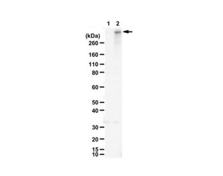
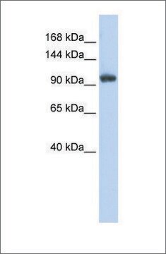
Western Blotting Analysis: 1 µg/mL from a representative lot detected endogenous HOIP (RNF31) in 60 µg of lysate from HEK293T and wild-type, but not HOIP-knockout, Jurkat cells, as well as the overexpression of an myc-tagged human HOIP in transfected HEK293T cells (Courtesy of Professor Kazuhiro Iwai, Kyoto University School of Medicine, Japan).

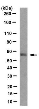
Western Blotting Analysis: 1 µg/mL from a representative lot detected endogenous HOIP (RNF31) in 60 µg of human (Jurkat, K562, U937), but not mouse (MEF & Bal17.2) cell lysates, as well as the overexpression of an myc-tagged human HOIP in transfected HEK293T cells (Courtesy of Professor Kazuhiro Iwai, Kyoto University School of Medicine, Japan).

Quality

Immunohistochemistry Analysis: A 1:10 dilution of this antibody detected HOIP in human adrenal gland tissue section.

Target description

~120 kDa observed. 119.7/57.63/102.5 kDa (isoform 1/2/3) calculated. Target band often appears as a doublet with the upper band representing ubiquitinated HOIP. Uncharacterized bands may be observed in some lysate(s).

Physical form

Format: Purified

Other Notes

Concentration: Please refer to lot specific datasheet.

Not finding the right product?  

Try our Product Selector Tool.

Storage Class Code

12 - Non Combustible Liquids

WGK

WGK 1

Flash Point(F)

Not applicable

Flash Point(C)

Not applicable


Certificates of Analysis (COA)

Search for Certificates of Analysis (COA) by entering the products Lot/Batch Number. Lot and Batch Numbers can be found on a product’s label following the words ‘Lot’ or ‘Batch’.

Already Own This Product?

Find documentation for the products that you have recently purchased in the Document Library.

Visit the Document Library

Our team of scientists has experience in all areas of research including Life Science, Material Science, Chemical Synthesis, Chromatography, Analytical and many others.

Contact Technical Service