Skip to Content
Merck
All Photos(2)

Key Documents

N5389

Sigma-Aldrich

Anti-Neurofilament H (200 kDa) Antibody

enhanced validation

mouse monoclonal, NE14

Synonym(s):

NF200 Antibody - Monoclonal Anti-Neurofilament 200 antibody produced in mouse, Neurofilament Antibody, Nf200 Antibody, Monoclonal Anti-Neurofilament heavy chain

Sign Into View Organizational & Contract Pricing


About This Item

MDL number:
UNSPSC Code:
12352203
NACRES:
NA.41

product name

Monoclonal Anti-Neurofilament 200 antibody produced in mouse, clone NE14, ascites fluid

biological source

mouse

Quality Level

conjugate

unconjugated

antibody form

ascites fluid

antibody product type

primary antibodies

clone

NE14, monoclonal

mol wt

antigen apparent mol wt 200 kDa

contains

15 mM sodium azide as preservative

species reactivity

pig, mouse, chicken, bovine, guinea pig, rabbit, rat, human

enhanced validation

independent ( Antibodies)
Learn more about Antibody Enhanced Validation

technique(s)

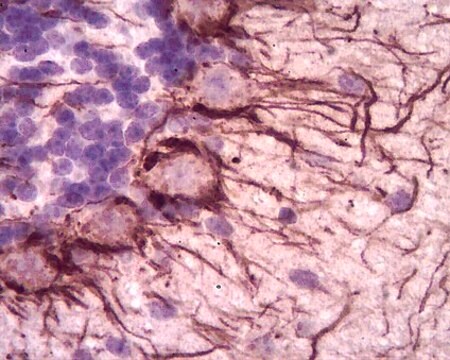
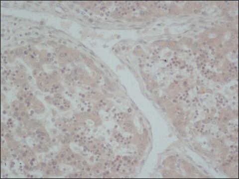
immunohistochemistry (formalin-fixed, paraffin-embedded sections): 1:40
immunohistochemistry (frozen sections): suitable
microarray: suitable
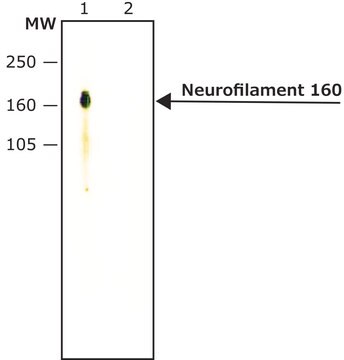
western blot: suitable

isotype

IgG1

UniProt accession no.

shipped in

dry ice

storage temp.

−20°C

target post-translational modification

unmodified

Gene Information

human ... NEFH(4744)

General description

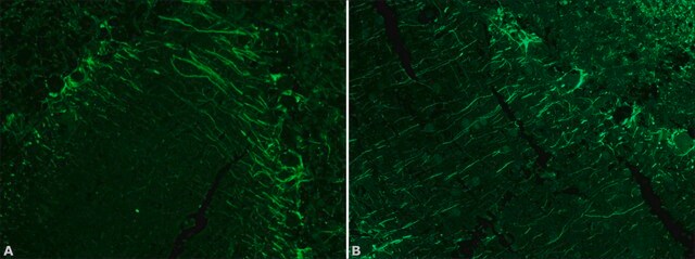
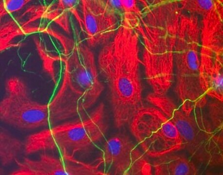
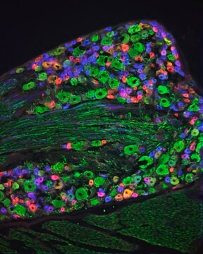
Monoclonal Anti-Neurofilament 200 (mouse IgG1 isotype) is derived from the hybridoma produced by the fusion of mouse myeloma cells and splenocytes from an immunized mouse. The neurofilaments are one of the five major groups of intermediate filaments (IFs) and are found predominantly in cells or tissues of neuronal origin. They are composed of three major proteins of apparent molecular weights 68 kD, 160 kD, and 200 kD and are named as NEFL (light), NEFM (medium) and NEFH (heavy) respectively. Neurofilament proteins are synthesized in the neuronal perikarya, assembled to form filaments and then slowly transported within the axons towards the synaptic terminals.

Immunogen

pig spinal cord.

Application

Applications in which this antibody has been used successfully, and the associated peer-reviewed papers, are given below.
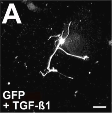
Immunofluorescence (1 paper)
Immunohistochemistry (1 paper)
Monoclonal Anti-Neurofilament 200 antibody produced in mouse has been used in
  • immunohistochemistry
  • immunolabeling
  • immunofluorescence

Biochem/physiol Actions

Neurofilament heavy chain (NEFH) gene was recently identified to cause autosomal dominant axonal Charcot-Marie-Tooth disease (CMT2cc). This gene plays a role in the pathogenesis of sporadic amyotrophic lateral sclerosis (ALS). The phosphorylation of neurofilament polypeptides has been suggested to modulate their function by influencing the interaction between neurofilament and cytoplasmic organelles.

Disclaimer

Unless otherwise stated in our catalog or other company documentation accompanying the product(s), our products are intended for research use only and are not to be used for any other purpose, which includes but is not limited to, unauthorized commercial uses, in vitro diagnostic uses, ex vivo or in vivo therapeutic uses or any type of consumption or application to humans or animals.

Not finding the right product?  

Try our Product Selector Tool.

Storage Class Code

10 - Combustible liquids

WGK

WGK 3

Flash Point(F)

Not applicable

Flash Point(C)

Not applicable


Certificates of Analysis (COA)

Search for Certificates of Analysis (COA) by entering the products Lot/Batch Number. Lot and Batch Numbers can be found on a product’s label following the words ‘Lot’ or ‘Batch’.

Already Own This Product?

Find documentation for the products that you have recently purchased in the Document Library.

Visit the Document Library

Cell cycle kinetics and immunohistochemical characterization of dissociated fetal neocortical cultures: evidence that differentiated neurons have mitotic capacity
Jacobs JS and Miller MW
Dev. Brain Res., 122(1), 67-80 (2000)
Neurofilament proteins are synthesized in nerve endings from squid brain
Crispino M, et al.
Journal of Neurochemistry, 61(3), 1144-1146 (1993)
Pamela Imperadore et al.
Scientific reports, 7, 46564-46564 (2017-04-21)
Regeneration is a process that restores structure and function of tissues damaged by injury or disease. In mammals complete regeneration is often unsuccessful, while most of the low phyla animals can re-grow many parts of their body after amputation. Cephalopod
C Lois et al.
Proceedings of the National Academy of Sciences of the United States of America, 90(5), 2074-2077 (1993-03-01)
Subventricular zone (SVZ) cells proliferate spontaneously in vivo in the telencephalon of adult mammals. Several studies suggest that SVZ cells do not differentiate after mitosis into neurons or glia but die. In the present work, we show that SVZ cells
Jon O Cleary et al.
NeuroImage, 56(3), 974-983 (2011-02-12)
Extensive worldwide efforts are underway to produce knockout mice for each of the ~25,000 mouse genes, which may give new insights into the underlying pathophysiology of neurological disease. Microscopic magnetic resonance imaging (μMRI) is a key method for non-invasive morphological

Our team of scientists has experience in all areas of research including Life Science, Material Science, Chemical Synthesis, Chromatography, Analytical and many others.

Contact Technical Service