Skip to Content
Merck
All Photos(4)

Documents

F3648

Sigma-Aldrich

Anti-Fibronectin antibody produced in rabbit

affinity isolated antibody, buffered aqueous solution

Synonym(s):

Fibronectin Antibody, Fibronectin Antibody - Anti-Fibronectin antibody produced in rabbit, Fibronectin Antibody Sigma

Sign Into View Organizational & Contract Pricing


About This Item

MDL number:
UNSPSC Code:
12352203
NACRES:
NA.23

biological source

rabbit

Quality Level

conjugate

unconjugated

antibody form

affinity isolated antibody

antibody product type

primary antibodies

clone

polyclonal

form

buffered aqueous solution

species reactivity

human

technique(s)

indirect ELISA: 1:10,000 using human fibronectin
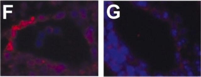
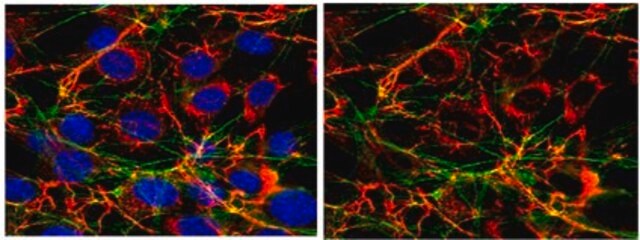
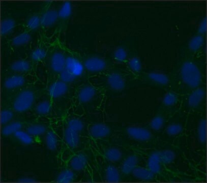
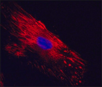
indirect immunofluorescence: 1:400 using human foreskin cultured fibroblasts
microarray: suitable
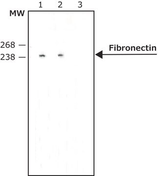
western blot: 1:1,000 using human plasma fibronectin

UniProt accession no.

application(s)

research pathology

shipped in

dry ice

storage temp.

−20°C

target post-translational modification

unmodified

Gene Information

human ... FN1(2335)

General description

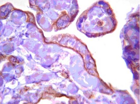
Antiserum is produced in rabbit using purified human fibronectin as the immunogen. Affinity isolated antibody is obtained from antiserum by immunospecific purification which removes essentially all rabbit serum proteins, including immunoglobulins, which do not specifically bind to human fibronectin. The antiserum is determined to be immunospecific for human fibronectin by immunofluorescent labeling of human fibroblast cell cultures, ELISA and immunoblotting. In immunoblotting, a specific band of fibronectin at 220 kDa is observed (another band at 94 kDa may be also be present) using human fibronectin. When used in immunoelectrophoresis, the antibody shows 1-2 arcs of precipitation versus normal human plasma. This product may be used for immunohistochemical localization of fibronectin in normal, inflamed and neoplastic tissues, for detection of fibronectin on cultured cells and structure and function studies of fibronectins in human and animal body fluids, tissues and cells. Affinity isolated antibody to human fibronectin can be used for immunofluorescent and immunoperoxidase staining of cultured cells, frozen sections and formalin fixed, paraffin-embedded tissues. Other fixatives, e.g. methacarn and ethanol, may also be used.

Fibronectin (FN) is composed of two nearly identical disulfide-bound polypeptides of molecular weight 220 kDa. Cellular fibronectin is structurally and antigenically similar to cold insoluble globulin from plasma, therefore polyclonal antibodies to either form usually cross-react. Careful analysis of the fibronectin molecule indicates that it contains several functionally and structurally distinct domains, which may bind to cell surfaces, collagen, fibrinogen or fibrin, complement, glycosaminoglycans, proteoglycans and heparin. Fibronectin 1 is encoded by the gene mapped to human chromosome 2q35.
Fibronectin (FN) is a multifunctional, extracellular matrix glycoprotein. Cellular fibronectin exists as dimeric or cross-linked multimeric forms, made up of fibroblasts, epithelial and other cell types. The dimer form composed of two nearly identical ~250 kDa subunits linked covalently to the C-termini by a pair of disulfide bonds. FN is present as dimeric form in plasma and secreted by hepatocytes.

Specificity

The product shows no cross-reaction with laminin, vitronectin, collagen type IV, or chondroitin sulfate types A, B and C using a dot blot immunoassay.

Immunogen

Fibronectin isolated from human plasma

Application

Anti-Fibronectin antibody is suitable for
  • immunohistochemistry (IHC) analysis to study the activity of pancreatic stellate cells in chronic pancreatitis
  • antibody blocking assays of bacterial cells to study the activity of fibronectin in the Yersinia pestis binding to host cells and Yop delivery
  • staining of endothelial cells as primary antibody
  • cell adhesion assays to study the effect of anti-human fibronectin antibodies on adhesion to endothelial cells
  • immunoprecipitation and western blot analysis of NF-κB dependent mammary tumorigenesis study
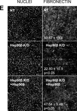
  • immunofluorescence staining to study the correlation between zyxin tails and fibrillar adhesions
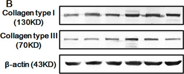
  • immunoblotting to study the expression levels of the pancreatic stellate cells (PSC) markers glial fibrillary acidic protein (GFAP), α-smooth muscle actin (α-SMA), collagen type I, and fibronectin in whole cell lysates
  • ELISA of fibronectin secretion
  • microarray analysis.

Biochem/physiol Actions

Fibronectin (FN) plays important roles in cell adhesion, migration, growth and differentiation. It has capacity to bind various functionally important domains such as heparin, collagen/gelatin, and fibrin. It also stimulates human non-small cell lung carcinoma.
Numerous studies have shown that fibronectin may enhance cell adhesion and spreading and affect the routes of cell migration both in vivo and in culture. Moreover, it has been shown that upon malignant transformation many cells lose most of their surface bound fibronectin. Fibronectin has been shown to also play a role in cellular morphology, cytoskeletal organization, phagocytosis, hemostasis, embryonic differentiation and wound repair. Fibronectin is produced by a wide variety of epithelial and mesenchymal cells in vitro including: fibroblasts, chondrocytes, myoblasts, Schwann cells, macrophages, hepatocytes and intestinal epithelial cells. Cellular fibronectin is present in many tissues including spleen, lymph node, tonsil, blood vessel walls, liver, kidney, muscle, skin, brain and peripheral nerves. It is found in basement membranes and in loose connective tissue stroma. It is also present in platelet α-granules and is expressed on the platelet surface after activation.

Physical form

Solution in 0.01M phosphate buffered saline, pH 7.4 containing 1% BSA and 15 mM sodium azide

Storage and Stability

For continuous use, store at 2-8°C for up to one month. For extended storage, the solution may be frozen in working aliquots. Repeated freezing and thawing, or storage in "frost-free" freezers, is not recommended. If slight turbidity occurs upon prolonged storage, clarify the solution by centrifugation before use.

Disclaimer

Unless otherwise stated in our catalog or other company documentation accompanying the product(s), our products are intended for research use only and are not to be used for any other purpose, which includes but is not limited to, unauthorized commercial uses, in vitro diagnostic uses, ex vivo or in vivo therapeutic uses or any type of consumption or application to humans or animals.

Not finding the right product?  

Try our Product Selector Tool.

Storage Class Code

10 - Combustible liquids

WGK

WGK 3

Flash Point(F)

Not applicable

Flash Point(C)

Not applicable

Personal Protective Equipment

dust mask type N95 (US), Eyeshields, Gloves

Certificates of Analysis (COA)

Search for Certificates of Analysis (COA) by entering the products Lot/Batch Number. Lot and Batch Numbers can be found on a product’s label following the words ‘Lot’ or ‘Batch’.

Already Own This Product?

Find documentation for the products that you have recently purchased in the Document Library.

Visit the Document Library

R P Casaroli Marano et al.
Investigative ophthalmology & visual science, 35(6), 2791-2803 (1994-05-01)
To examine the possible role of some adhesion multifunctional glycoproteins of the extracellular matrix, such as fibronectin (FN), laminin (LN), vitronectin (VN) and their receptors (beta 1-subunit complex and alpha v beta 3 integrins) in events of cell migration and
Tsutomu Inoue et al.
Biochemical and biophysical research communications, 517(1), 96-102 (2019-07-20)
Here we evaluated the efficacy of depleting cellular communication network factor 2 (CCN2) produced by renal tubular epithelial cells in preventing the progression of severe acute kidney injury (AKI) to chronic kidney disease (CKD). We used conditional Ccn2 knockout mice
Christoph W Michalski et al.
Journal of translational medicine, 5, 63-63 (2007-12-07)
Interactions between mononuclear cells and activated pancreatic myofibroblasts (pancreatic stellate cells; PSC) may contribute to inflammation and fibrosis in chronic pancreatitis (CP). Markers of fibrosis and inflammation were concomitantly analysed by immunohistochemistry in chronic pancreatitis tissues. In vitro, PSC were
Molecular identification of COL6A3-CSF1 fusion transcripts in tenosynovial giant cell tumors.
Moller E, et al.
Genes Chromosomes Cancer, 47(1), 21-25 (2008)
Fibronectin molecule visualized in electron microscopy: a long, thin, flexible strand.
Erickson H P, et al.
The Journal of Cell Biology, 91(3), 673-678 (1981)

Our team of scientists has experience in all areas of research including Life Science, Material Science, Chemical Synthesis, Chromatography, Analytical and many others.

Contact Technical Service