05316
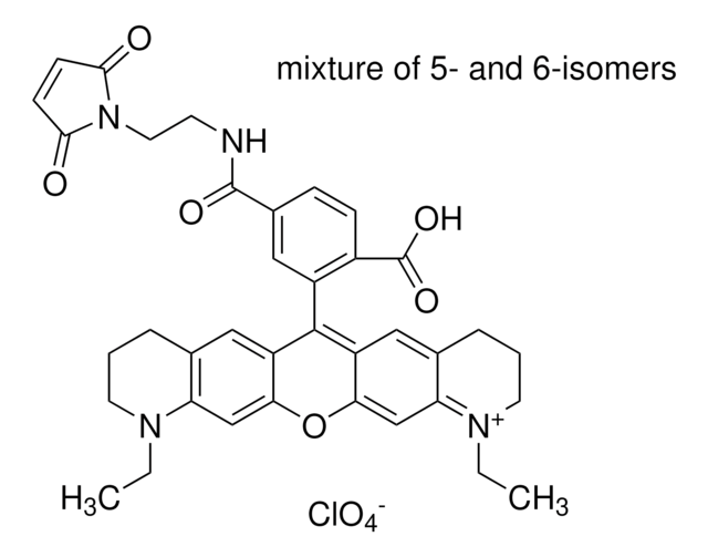
Atto 647N maleimide
BioReagent, suitable for fluorescence, ≥90% (HPLC)
Sign Into View Organizational & Contract Pricing
All Photos(1)
About This Item
Recommended Products
product line
BioReagent
Assay
≥90% (HPLC)
≥90% (degree of coupling)
manufacturer/tradename
ATTO-TEC GmbH
λ
in ethanol (with 0.1% trifluoroacetic acid)
UV absorption
λ: 640-646 nm Amax
suitability
suitable for fluorescence
detection method
fluorometric
storage temp.
−20°C
Related Categories
Application
- Preparation of homogeneous samples of double-labelled protein suitable for single-molecule FRET measurements.: This study explores the use of Atto 647N maleimide for the preparation of homogeneously double-labelled protein samples, facilitating precise single-molecule FRET measurements to analyze protein dynamics and interactions (Lerner et al., 2013).
Legal Information
This product is for Research use only. In case of intended commercialization, please contact the IP-holder (ATTO-TEC GmbH, Germany) for licensing.
Storage Class Code
11 - Combustible Solids
WGK
WGK 3
Flash Point(F)
Not applicable
Flash Point(C)
Not applicable
Personal Protective Equipment
dust mask type N95 (US), Eyeshields, Gloves
Choose from one of the most recent versions:
Already Own This Product?
Find documentation for the products that you have recently purchased in the Document Library.
Customers Also Viewed
STED microscopy to monitor agglomeration of silica particles inside A549 cells.
Schubbe, S., et al.
Advanced Engineering Materials, 12, 417-422 (2010)
A novel nanoscopic tool by combining AFM with STED microscopy.
Harke, B., et al.
Optical Nanoscopy, 1, 3-3 (2012)
Volker Westphal et al.
Science (New York, N.Y.), 320(5873), 246-249 (2008-02-23)
We present video-rate (28 frames per second) far-field optical imaging with a focal spot size of 62 nanometers in living cells. Fluorescently labeled synaptic vesicles inside the axons of cultured neurons were recorded with stimulated emission depletion (STED) microscopy in
Marisa L Martin-Fernandez et al.
International journal of molecular sciences, 13(11), 14742-14765 (2012-12-04)
Insights from single-molecule tracking in mammalian cells have the potential to greatly contribute to our understanding of the dynamic behavior of many protein families and networks which are key therapeutic targets of the pharmaceutical industry. This is particularly so at
S E D Webb et al.
Optics express, 16(25), 20258-20265 (2008-12-10)
We combine single molecule fluorescence orientation imaging with single-pair fluorescence resonance energy transfer microscopy, using a total internal reflection microscope. We show how angles and FRET efficiencies can be determined for membrane proteins at the single molecule level and provide
Our team of scientists has experience in all areas of research including Life Science, Material Science, Chemical Synthesis, Chromatography, Analytical and many others.
Contact Technical Service


