Skip to Content
Merck
All Photos(4)

Key Documents

MABT1354

Sigma-Aldrich

Anti-CP110 Antibody, clone 140-195-5

clone 140-195-5, from mouse

Synonym(s):

Centriolar coiled-coil protein of 110 kDa, Centrosomal protein of 110 kDa, Cep110

Sign Into View Organizational & Contract Pricing


About This Item

UNSPSC Code:
12352203
eCl@ss:
32160702
NACRES:
NA.41

biological source

mouse

Quality Level

antibody form

purified immunoglobulin

antibody product type

primary antibodies

clone

140-195-5, monoclonal

species reactivity

human

technique(s)

immunocytochemistry: suitable
immunofluorescence: suitable
immunoprecipitation (IP): suitable
western blot: suitable

isotype

IgG1κ

NCBI accession no.

UniProt accession no.

shipped in

ambient

target post-translational modification

unmodified

Gene Information

human ... CCP110(9738)

General description

Centriolar coiled-coil protein of 110 kDa (UniProt: O43303; also known as Centrosomal protein of 110 kDa, CP110, Cep110) is encoded by the CCP110 (also known as CEP110, CP110, KIAA0419) gene (Gene ID: 9738) in human. CP110 is a cytoplasmic protein that is essential for centrosome duplication at different stages of procentriole formation. It acts as a key negative regulator of ciliogenesis in collaboration with CEP97 by capping the mother centriole thereby preventing cilia formation. It is also involved in the assembly of the mother centriole subdistal appendages thereby affecting the fusion of recycling endosomes to basal bodies during cilia formation. Two isoforms of CP110 have been reported that are produced by alternative splicing. CP110 is highly expressed in testis and intermediate level of expression is observed in spleen, thymus, prostate, small intestine, colon and peripheral blood leukocytes. CP110 levels re up-regulated during the transition from G1 to S phase of the cell cycle and highest levels are reported in S phase, following which the levels decline significantly. During the G2 phase it is ubiquitinated that leads to its proteasomal degradation, which prevents centrosome reduplication. And, de-ubiquitinated by USP33 in S and G2/M phase stabilizes CCP110 during the period when centrioles duplicate and elongate.

Specificity

Clone 140-195-5 detects Centriolar coiled-coil protein of 110 kDa (CP110) in human cells., It targets an epitope within the first 149 amino acids from the N-termina region.

Immunogen

Epitope: N-terminus
His-tagged recombinant fragment corresponding to the first 149 amino acids from the N-terminal region of human Centriolar coiled-coil protein of 110 kDa.

Application

Anti-CP110, clone 140-195-5 , Cat. No. MABT1354, is a mouse monoclonal antibody that detects Centriolar coiled-coil protein of 110 kDa and has been tested for use in immunocytochemistry, Immunofluorescence, Immunoprecipitation, and Western Blotting.
Immunoprecipitation Analysis: A representative lot detected CP110 in HEK293T cell lysate (Courtesy of Prof. Erich A. Nigg, Biozentrum, University of Basel, Switzerland).

Immunocytochemistry Analysis: A 1:50 dilution from a representative lot detected CP110 in U2OS cell line.

Western Blotting Analysis: A representative lot detected CP110 in Western Blotting applications (Bauer, M., et. al. (2016). EMBO J. 35(19):2152-2166).

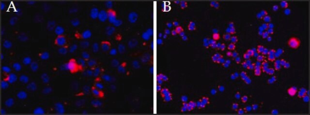
Immunofluorescence Analysis: A representative lot detected CP110 in U20S cells (Courtesy of Prof. Erich A. Nigg, Biozentrum, University of Basel, Switzerland).

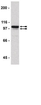
Western Blotting Analysis: A representative lot detected CP110 in 293T cell lysate (Courtesy of Prof. Erich A. Nigg, Biozentrum, University of Basel, Switzerland).

Immunofluorescence Analysis: A representative lot detected CP110 in Western Blotting applications (Bauer, M., et. al. (2016). EMBO J. 35(19):2152-2166; Arquint, C., et. al. (2014). Curr Biol. 24(4):351-60).
Research Category
Cell Structure

Quality

Evaluated by Western Blotting in HEK293 cell lysate.

Western Blotting Analysis: A 1:250 dilution of this antibody detected CP110 in HEK293 cell lysate.

Target description

~150 kDa observed; 113.42 kDa calculated. Uncharacterized bands may be observed in some lysate(s).

Physical form

Format: Purified
Protein G purified
Purified mouse monoclonal antibody IgG1 in buffer containing 0.1 M Tris-Glycine (pH 7.4), 150 mM NaCl with 0.05% sodium azide.

Storage and Stability

Stable for 1 year at 2-8°C from date of receipt.

Other Notes

Concentration: Please refer to lot specific datasheet.

Disclaimer

Unless otherwise stated in our catalog or other company documentation accompanying the product(s), our products are intended for research use only and are not to be used for any other purpose, which includes but is not limited to, unauthorized commercial uses, in vitro diagnostic uses, ex vivo or in vivo therapeutic uses or any type of consumption or application to humans or animals.

Not finding the right product?  

Try our Product Selector Tool.

Storage Class Code

12 - Non Combustible Liquids

WGK

WGK 1

Flash Point(F)

Not applicable

Flash Point(C)

Not applicable


Certificates of Analysis (COA)

Search for Certificates of Analysis (COA) by entering the products Lot/Batch Number. Lot and Batch Numbers can be found on a product’s label following the words ‘Lot’ or ‘Batch’.

Already Own This Product?

Find documentation for the products that you have recently purchased in the Document Library.

Visit the Document Library

Our team of scientists has experience in all areas of research including Life Science, Material Science, Chemical Synthesis, Chromatography, Analytical and many others.

Contact Technical Service