Prestige Antibodies® on The Cell Atlas
In 2015, the Human Protein Atlas project presented a complete map of the human proteome expression in human tissues. The Tissue Atlas and Cancer Atlas includes data for 17,500 Prestige Antibodies®.
The newest addition to the Human Protein Atlas is the Cell Atlas - a fantastic tool for anyone working with antibodies for ICC-IF or in cell biology in general. The Cell Atlas presents data for more than 10,000 Prestige Antibodies®.
Biological processes within the cell are fundamental to all life. A key to better understand the mechanisms of living cells is to determine the subcellular localization of proteins active in the cells. Different organelles offer environment with different physiological conditions, constituents and interaction partners, which determine protein function.
The Cell Atlas part of the Human Protein Atlas visualizes the spatial distribution of proteins in the human cell. For each protein there are immunofluorescent stainings from target specific antibodies in several human cell lines, annotated subcellular locations and target gene RNA expression in a vast number of cell lines. For many of the proteins, there are also results of different validation assays performed, such as siRNA knockdown, GFP co-localization and cell cycle dependency analyses.

Screen shot from the Human Protein Atlas showing part of the information for Cyclin B1 on the Cell Atlas. Cyclin B1 is localized in the Cytosol of the cell
The Cell Atlas is divided in four different chapters, providing knowledge-based analysis of the human cellular proteomes:
The different chapters include descriptions and explorations of protein expression patterns, a complete list of proteins expressed in the proteome, as well as examples of detailed images illustrating the subcellular spatial distribution patterns.

Image courtesy of the Human Protein Atlas
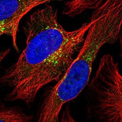
For the Cell Atlas, confocal microscopy has been used to provide high-resolution, four-color images in cell lines. Three different organelle markers are displayed together with the specific antibody as different channels in the multicolor images:
- Antibody stained in green
- Nucleus stained in blue
- Microtubules in red
- ER in yellow
By using the "toggle channels"-button on the high-resolution images, the different channels can be turned on and off.


Immunofluorescence of U-2 OS cell line. Antibody HPA030741 (Anti-CCNB1) in green, nucleus stained in blue and microtubules in red. In the right image, only the green channel, showing the antibody staining, is turned on.
The images in the Cell Atlas provide single cell resolution, which makes it possible to study variations in protein expression patterns from cell to cell. The cells are grown under asynchronous conditions making the most likely explanation for single cell variations to be an effect of the cell cycle.

The Anti-MRPS7, HPA022522 antibody (stained in green) shows single cell variations in A-431 cell line
Below are example antibodies of Prestige Antibodies® highlighting three different organelles.
The Anti-DDX39B antibody (HPA058450) detects the spliceosome RNA helicase DDX39B protein that is involved in nuclear export of spliced and unspliced mRNA. In the human cell line MCF7, immunofluorescent staining using the HPA058450 antibody shows localization to nuclear speckles (antibody staining in green).

The Anti-COPG1 antibody (HPA037866) is designed against the Coatomer subunit gamma-1 protein of the coatamer complex that functions in mediating protein transport from the ER, via the Golgi up to the trans Golgi network. The HPA037866 antibody shows localization to the Golgi apparatus by immunofluorescent staining of human cell line U-2 OS (antibody staining in green).

The Anti-KIF5B antibody (HPA037589) detects the Kinesin-1 heavy chain protein that is a microtubule-dependent motor required for normal distribution of mitochondria and lysosomes. Using immunofluorescence of human cell line U-2 OS, the HPA037589 antibody shows localization to cytosol and microtubule organizing center (antibody staining in green).

All antibodies developed for the Human Protein Atlas, that consists of the Cell Atlas, the Tissue Atlas and the Cancer Atlas, are available from us as Prestige Antibodies®.
To continue reading please sign in or create an account.
Don't Have An Account?