Live Cell Fluorescent Organelle Dyes and Stains
Eukaryotic cells contain subcellular structures responsible for carrying out essential functions for cell survival, including generating energy (ATP), synthesizing and trafficking proteins, and cell waste removal. Due to their specialized functions and well-defined nature, it is often useful in cell biology studies to label key organelles, such as the cell membrane, nucleus, cytoplasm, mitochondria, lysosomes, endoplasmic reticulum (ER), Golgi apparatus, and cytoskeleton proteins (e.g., actin). More specifically, utilizing organelle dyes as counterstains in live cell imaging techniques has proven to be invaluable in the study of cellular function. We offer many fluorescent dyes and markers highly specific for diverse organelles that can be used to monitor cell health, cell death, metabolic activity, autophagy, cell tracking,and cell migration and invasion.
Mitochondria Dyes
Mitochondria are double-membrane-bound organelles, responsible for cellular respiration and the production of energy as adenosine triphosphate (ATP). Mitochondria are also involved in other tasks, such as cell signaling, cellular differentiation, and cell death—as well as maintaining control of the cell cycle and growth. Dependent on membrane potential, fluorescent live cell dyes for mitochondria can be used to analyze overall cell health and vitality, in addition to mitochondrial activity.
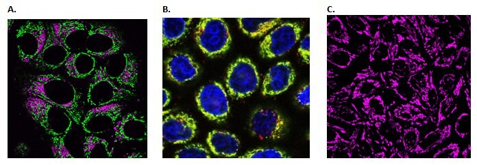
Figure 1. Live cell imaging of cellular mitochondria.A) Live HeLa cells co-stained with BioTracker NIR650 Lysosome Dye (SCT139) and BioTracker 488 Green Mitochondria Dye (SCT136). B) Live HeLa cells co-stained with BioTracker ATP-Red live cell dye (SCT045), Hoechst (94403) and a green mitochondria-specific dye showing localization within mitochondria. C) Live HeLa cells stained with BioTracker 633 Red Mitochondria Dye (SCT137).
Lysosome Dyes
Lysosomes are membrane-bound organelles that act as the waste disposal system of the cell. As highly acidic (pH 4-5) organelles, lysosomes contain a variety of enzymes capable of breaking down various biomolecules including peptides, nucleic acids, carbohydrates, and lipids. In addition to biomolecule dissolution, lysosomes are also involved in autophagy and apoptosis. Dependent on the internal acidic environment, live cell fluorescent dyes for lysosomes can be used to analyze lysosomal activity, autophagy events, vesicle trafficking, and overall cell health.
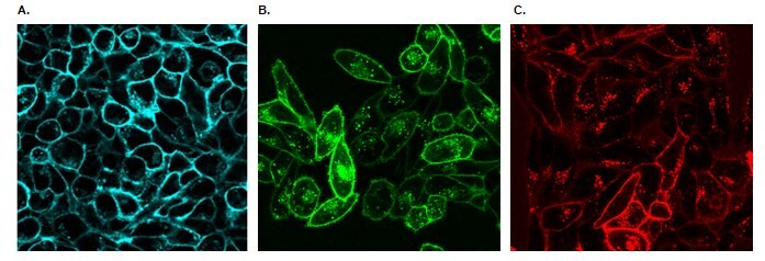
Figure 2. Live cell imaging of cellular lysosomes.A) HeLa cells expressing Mitochondria-GFP were stained with BioTracker 560 Orange Lysosome Dye (SCT019) and Hoechst DNA stain (Blue). B) Live HeLa cells stained with Live Cell Microtubule Stain (Blue) and BioTracker 540 Red Lysosome Dye (SCT141). C) Live Hela cells stained with BioTracker 540 Red Lysosome Dye (SCT141).
Nuclear Dyes
Containing nearly all of the cell's genetic material, the cell nucleus is organized in chromosomes, formed from the complexation of long linear DNA with proteins. Live cell nuclear stains can be used either for cell tracking, or as counterstains with other fluorescent dyes or cell reporters. We offer dyes with several color options, including long-wavelength, far-red nuclear dyes. In comparison to traditional UV excitable DNA dyes (e.g., Hoechst 33342), long-wavelength nuclear dyes demonstrate reduced cellular toxicity during live cell imaging experiments.
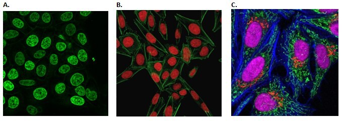
Figure 3. Live cell imaging of the cellular nucleus.A) Live HeLa cells stained with BioTracker 488 Green Nuclear Dye (SCT120). B) Live cell staining of HeLa cells using the BioTracker NIR694 Nuclear Dye (water-based) and FITC-phalloidin. C) Fixed cell staining of HeLa cells using BioTracker NIR694 Nuclear Dye (in DMSO), BioTracker 488 Green Mitochondria Dye and CF405-phalloidin.
Cell Membrane Dyes
Protecting and sequestering the cell interior, the cell membrane is composed primarily of lipids and proteins. In addition to controlling movement across the bilayer, cell membranes are involved in a variety of cellular processes such as cell adhesion, ion conductivity, cell signaling, and extracellular structure attachment. BioTracker cytoplasmic membrane dyes are lipophilic carbocyanines capable of labeling cell membranes in a stable and non-toxic manner. Due to their low cytotoxicity, these dyes are suitable for long-term cell tracking and imaging. Unlike PKH dyes, BioTracker cytoplasmic membrane dyes are ready-to-use and do not require a complicated hypo-osmotic labeling protocol. BioTracker dyes can be added directly to normal culture media to label either suspended or adherent cells.

Figure 4. Live cell imaging of cellular membranes.A) Live HeLa cells stained with BioTracker 400 Blue Cytoplasmic Membrane Dye (SCT109). B) Live cell staining of Hela cells using BioTracker 490 Green Cytoplasmic Membrane Dye (SCT106). C) Live cell staining of Hela cells using BioTracker NIR750 Cytoplasmic Membrane Dye (SCT113).
Cytoskeleton Dyes
As a complex network of interlinking filaments and microtubules extending throughout the cytoplasm, the cytoskeleton provides structural support to the cell and controls many processes, including endocytosis, cytokinesis, cell migration, invasion, and movement. In contrast to traditional, fixed F-actin stains (e.g., conjugated phalloidin dyes), BioTracker microtubule cytoskeleton dyes are cell-permeable paclitaxel probes that can be imaged without a wash step. Offered in green and red fluorescent color options, these simple, rapid and sensitive stains can be used to monitor cytoskeleton dynamics in real time.
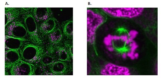
Figure 5. Live cell imaging of the cytoskeleton.A) Live HeLa cells stained with BioTracker 488 Green Microtubule Cytoskeleton Dye (SCT142) and BioTracker NIR650 Lysosome Dye (SCT139). B) Mitotic spindle stained with BioTracker 488 Green Microtubule Cytoskeleton Dye (SCT142). DNA is stained with BioTracker NIR694 Nuclear Dye (water-based) (SCT117).
Lipid Dyes
As fundamental building blocks of the cell, lipids serve many important cellular functions but have also been implicated in various diseases, including cardiovascular and neurological disorders. Intracellular lipid droplets are cytoplasmic organelles involved in the storage and regulation of triglycerides and cholesterol esters. BioTracker lipid dye rapidly stains lipid droplets in either live or fixed cells without any wash steps and with minimal background staining of cellular membranes or other organelles. Live cells may subsequently fixed and permeabilized after staining with BioTracker lipid droplet dye.
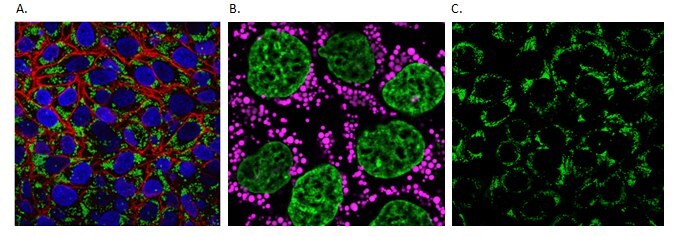
Figure 6. Live cell imaging of cellular lipids.A) Live HeLa cells stained with BioTracker 488 Green Lipid Dye (SCT144), Hoechst (94403) and BioTracker 655 Red Cytoplasmic Membrane Dye (SCT108). B) HeLa cells stained with BioTracker 610 Red Lipid Droplet Dye (magenta) and BioTracker 488 Green Nuclear Dye (SCT120). C) HeLa cells stained with BioTracker 488 Green Lipid Dye (SCT144).
Conclusion
Restricting cell culture imaging techniques to fixed tissues and cells limits the information a researcher can obtain about the system of interest, as the observer is only able to capture a moment in cellular time. Our live cell fluorescent organelle dyes permit the selective staining of specific organelles without increased cytotoxicity. Organelle staining in live cells can help researchers to confirm the identity of specific proteins or targets within cells and provide a better understanding of cellular processes. Our broad portfolio ensures we have the organelle-specific dye in the color you need for your live cell imaging experiments.
To continue reading please sign in or create an account.
Don't Have An Account?