G7777
Anti-Mouse IgG (whole molecule)–Gold antibody produced in goat
affinity isolated antibody, aqueous glycerol suspension, 10 nm (colloidal gold)
Sign Into View Organizational & Contract Pricing
All Photos(1)
About This Item
Recommended Products
biological source
goat
Quality Level
conjugate
gold conjugate
antibody form
affinity isolated antibody
antibody product type
secondary antibodies
clone
polyclonal
form
aqueous glycerol suspension
particle size
10 nm (colloidal gold)
storage temp.
2-8°C
target post-translational modification
unmodified
Looking for similar products? Visit Product Comparison Guide
General description
Immunoglobulin G (IgG) is a glycoprotein antibody that regulates immune responses such as phagocytosis and is also involved in the development of autoimmune diseases . Mouse IgGs have four distinct isotypes, namely, IgG1, IgG2a, IgG2b, and IgG3. IgG1 regulates complement fixation in mice . Anti-Mouse IgG (whole molecule)-Gold antibody is specific for mouse IgG.
Immunogen
Purified mouse IgG
Application
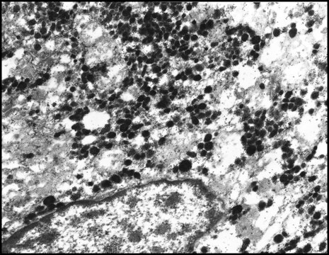
Anti-Mouse IgG (whole molecule)-Gold antibody is suitable for use in immunoelectron microscopy .
Other Notes
Antibody adsorbed with human serum proteins.
Physical form
Conjugate is suspended in Tris buffered saline, pH 8.2, containing 1% (w/v) BSA, 30% (v/v) glycerol and 15 mM sodium azide.
Disclaimer
Unless otherwise stated in our catalog or other company documentation accompanying the product(s), our products are intended for research use only and are not to be used for any other purpose, which includes but is not limited to, unauthorized commercial uses, in vitro diagnostic uses, ex vivo or in vivo therapeutic uses or any type of consumption or application to humans or animals.
Not finding the right product?
Try our Product Selector Tool.
Storage Class Code
12 - Non Combustible Liquids
WGK
WGK 2
Flash Point(F)
Not applicable
Flash Point(C)
Not applicable
Personal Protective Equipment
dust mask type N95 (US), Eyeshields, Gloves
Choose from one of the most recent versions:
Already Own This Product?
Find documentation for the products that you have recently purchased in the Document Library.
Chongluo Fu et al.
Diseases of aquatic organisms, 65(1), 17-22 (2005-07-27)
Recently, an enveloped, spherical RNA virus was identified as the causative agent of mass mortalities among adult scallop Chlamys farreri, which is cultured on the northern coast of China. Hybridomas were prepared from mice immunized with highly purified virions. Four
S B Shah et al.
Chromosoma, 105(2), 111-121 (1996-08-01)
Immunoelectron microscopy with anti-nucleolin defined substructures within the multiple nucleoli of biosynthetically active stage II-III oocytes and within the nucleoli of relatively quiescent stage VI oocytes of Xenopus laevis. Dense fibrillar components (DFCs) of nucleoli from stage II-III oocytes consisted
Maria Gregori et al.
Nanomedicine : nanotechnology, biology, and medicine, 13(2), 723-732 (2016-11-05)
Aggregation of amyloid-β peptide (Aβ) is a key event in the pathogenesis of Alzheimer's disease (AD). We investigated the effects of nanoliposomes decorated with the retro-inverso peptide RI-OR2-TAT (Ac-rGffvlkGrrrrqrrkkrGy-NH2) on the aggregation and toxicity of Aβ. Remarkably low concentrations of
William D Gilliland et al.
PloS one, 4(10), e7544-e7544 (2009-10-23)
The protein kinases Mps1 and Polo, which are required for proper cell cycle regulation in meiosis and mitosis, localize to numerous ooplasmic filaments during prometaphase in Drosophila oocytes. These filaments first appear throughout the oocyte at the end of prophase
Xiaoman Zhang et al.
PloS one, 17(6), e0270634-e0270634 (2022-06-25)
Extracellular vesicles (EVs) have attracted much attention as potential diagnostic biomarkers for human diseases. Although both plasma and serum are utilized as a source of blood EVs, it remains unclear whether, how and to what extent the choice of plasma
Our team of scientists has experience in all areas of research including Life Science, Material Science, Chemical Synthesis, Chromatography, Analytical and many others.
Contact Technical Service


