Skip to Content
Merck
All Photos(1)

Key Documents

MABT1569

Sigma-Aldrich

Anti-Dystrophin MANEX4850E Antibody, clone 8C5

Synonym(s):

DMD

Sign Into View Organizational & Contract Pricing


About This Item

UNSPSC Code:
12352203
NACRES:
NA.46
clone:
8C5, monoclonal
application:
IF
IHC
WB
species reactivity:
mouse, human
technique(s):
immunofluorescence: suitable
immunohistochemistry: suitable
western blot: suitable
citations:

biological source

mouse

Quality Level

antibody form

purified antibody

antibody product type

primary antibodies

clone

8C5, monoclonal

mol wt

calculated mol wt 426.75 kDa
observed mol wt ~430 kDa

purified by

using protein G

species reactivity

mouse, human

packaging

antibody small pack of 100

technique(s)

immunofluorescence: suitable
immunohistochemistry: suitable
western blot: suitable

isotype

IgG2aκ

epitope sequence

Internal

Protein ID accession no.

UniProt accession no.

storage temp.

2-8°C

Gene Information

human ... DMD(1756)

Specificity

Clone 8C5 is a mouse monoclonal antibody that detects the deletion prone region of Dystrophin. It targets an epitope within 295 amino acids from the internal region.

Immunogen

Recombinant human dystrophin fragment corresponding to amino acids 2145-2439 encoded by exons 45-50; as β-galactosidase fusion protein.

Application

Quality Control Testing

Evaluated by Western Blotting in Human muscle tissue extract.

Western Blotting Analysis: A 1:1,000 dilution of this antibody detected Dystrophin in Human muscle tissue extract, but not in HeLa cell lysate.

Tested Applications

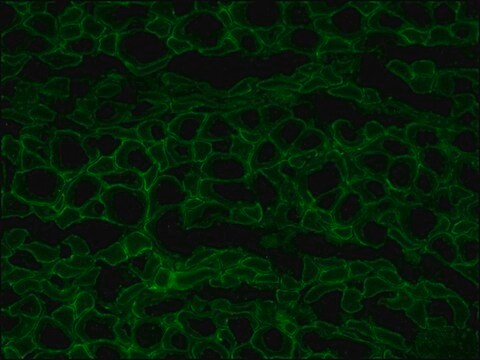
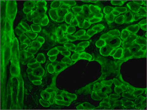
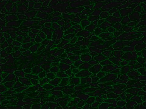
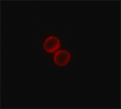
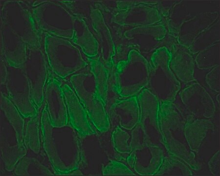
Immunofluorescence Analysis: A representative lot detected Dystrophin in Immunofluorescence applications (Lim, K.R.Q., et al. (2022). Proc Natl Acad Sci USA. 119(9): e2112546119).

Western Blotting Analysis: A representative lot detected Dystrophin in Western Blotting applications (Lim, K.R.Q., et al. (2022). Proc Natl Acad Sci USA. 119(9): e2112546119).

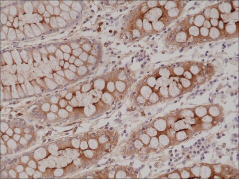
Immunohistochemistry Applications: A representative lot detected Dystrophin in Immunohistochemistry applications (Lim, K.R.Q., et al. (2022). Proc Natl Acad Sci USA. 119(9) e2112546119).

Note: Actual optimal working dilutions must be determined by end user as specimens, and experimental conditions may vary with the end user.

Target description

Dystrophin (UniProt: P11532; also known as DMD) is encoded by the DMD gene (Gene ID: 1756) in human. Duchenne muscular dystrophy is caused by mutations in DMD gene. Most subjects have out-of-frame deletions in the DMD gene that leads to lack of dystrophin in muscle. Dystrophin is a rod-shaped cytoplasmic protein and a vital part of a protein complex that connects the cytoskeleton of a muscle fiber to the surrounding extracellular matrix through the cell membrane. It is expressed in muscle fibers accumulating in the costameres of myoplasm at the sarcolemma. Its expression is also reported in brain, muscle, kidney, lung, and testis. Dystrophin is localized to the inner part of the muscle fiber cell membrane (sarcolemma), where it forms the dystrophin-associated glycoprotein complex (DGC) that links the extracellular matrix to the actin cytoskeleton. The N-terminus is the actin-binding domain, and the carboxy-terminal domain interacts with β-dystroglycan as well as dystrobrevin and the syntrophin. The central rod domain that comprises the major mass of the dystrophin molecule forms a flexible, rod-shaped structure. Dystrophin plays an important role in stabilizing the muscle fiber against the mechanical forces of muscle contraction by providing a shock-absorbing connection between the cytoskeleton and the extracellular matrix. The DMD gene is the largest known gene in humans and mutations in this gene are known to cause Duchenne muscular dystrophy, a sex-linked recessive disorder that affects males aged 3 to 7 year as proximal muscle weakness with steady progression. Clone 8C5 is generated from the deletion-prone region of the Duchenne muscular dystrophy gene. MANEX4850E corresponds to exons 48 to 50. Exon skipping studies have shown that phosphorodiamidate morpholino oligomers (PMOs) can convert out of-frame to in-frame mutations, which produce truncated, partially functional dystrophin. Seventeen different isoforms of Dystrophin have been described that are produced by alternative promoter usage and alternative splicing. (Ref.: Lim, KRQ., et al. (2022). Proc. Natl. Acad. Sci. USA. 119(9); e2112546119; Thanh, LT., et al. (1995). Am. J. Med. Genet. 58(2); 177-186).

Physical form

Purified mouse monoclonal antibody IgG2a in buffer containing 0.1 M Tris-Glycine (pH 7.4), 150 mM NaCl with 0.05% sodium azide.

Reconstitution

1.0 mg/mL. Please refer to guidance on suggested starting dilutions and/or titers per application and sample type.

Storage and Stability

Recommended storage: +2°C to +8°C.

Other Notes

Concentration: Please refer to the Certificate of Analysis for the lot-specific concentration.

Disclaimer

Unless otherwise stated in our catalog or other company documentation accompanying the product(s), our products are intended for research use only and are not to be used for any other purpose, which includes but is not limited to, unauthorized commercial uses, in vitro diagnostic uses, ex vivo or in vivo therapeutic uses or any type of consumption or application to humans or animals.

Not finding the right product?  

Try our Product Selector Tool.

Storage Class Code

12 - Non Combustible Liquids

WGK

WGK 1

Flash Point(F)

Not applicable

Flash Point(C)

Not applicable


Certificates of Analysis (COA)

Search for Certificates of Analysis (COA) by entering the products Lot/Batch Number. Lot and Batch Numbers can be found on a product’s label following the words ‘Lot’ or ‘Batch’.

Already Own This Product?

Find documentation for the products that you have recently purchased in the Document Library.

Visit the Document Library

Our team of scientists has experience in all areas of research including Life Science, Material Science, Chemical Synthesis, Chromatography, Analytical and many others.

Contact Technical Service