Skip to Content
Merck
All Photos(1)

Key Documents

05-362

Sigma-Aldrich

Anti-Cyclin D1/2 Antibody, clone 5D4

clone 5D4, Upstate®, from mouse

Sign Into View Organizational & Contract Pricing


About This Item

UNSPSC Code:
12352203
eCl@ss:
32160702
NACRES:
NA.41

biological source

mouse

Quality Level

antibody form

purified antibody

antibody product type

primary antibodies

clone

5D4, monoclonal

species reactivity

rat, mouse, human, hamster

manufacturer/tradename

Upstate®

technique(s)

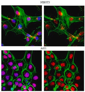
immunocytochemistry: suitable
immunoprecipitation (IP): suitable
western blot: suitable

isotype

IgG2a

NCBI accession no.

UniProt accession no.

shipped in

dry ice

target post-translational modification

unmodified

Gene Information

human ... CCND1(595)

General description

During each cell cycle cyclins undergo periodic accumulation and destruction. As key regulators of the cell cycle the cyclins control important transitions by acting as regulatory subunits of the Cdks. Early in the G1 phase of the cell cycle, cyclin D1 induction is followed by cyclin E induction. This sequential progression is marked early on in G1 by the activation of Cdk4 and in mid to late G1 by the activation of Cdk2 and the hyperphosphorylation of pRB. The final transition into S phase is thought to be dependent on the increased expression and association of cyclin E and Cdk2. Cyclin D2 is also a G1 cyclin required for G1 phase progression and is a strong candidate for a proto oncogene. Cyclin D2 can phosphorylate pRB when associated with cdk 4 and / or cdk 6.

Specificity

Recognizes p36 cyclin D1 and p34 cyclin D2; no cross-reactivity with cyclin A, B or D3.

Immunogen

Recombinant human cyclin D1

Application

Anti-Cyclin D1/2 Antibody, clone 5D4 is a high quality Mouse Monoclonal Antibody for the detection of Cyclin D1/2 & has been validated in ICC, IP & WB.
Research Category
Epigenetics & Nuclear Function
Research Sub Category
Cell Cycle, DNA Replication & Repair

Quality

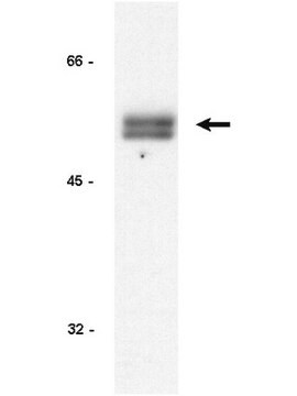
routinely evaluated by immunoblot on RIPA lysate from human A431 cells

Target description

34/36 kDa

Linkage

Replaces: 04-1151

Physical form

Format: Purified
Protein A purified
Protein A purified IgG2a in 50% storage buffer containing PBS, pH 7.2 and 50% glycerol.

Storage and Stability

Maintain for 2 years at -20°C from date of shipment. Aliquot to avoid repeated freezing and thawing. For maximum recovery of product, centrifuge the original vial after thawing and prior to removing the cap.

Analysis Note

Control
Positive Antigen Control: Catalog #12-301, non-stimulated A431 cell lysate. Add 2.5µL of 2-mercaptoethanol/100µL of lysate and boil for 5 minutes to reduce the preparation. Load 20µg of reduced lysate per lane for minigels.

Legal Information

UPSTATE is a registered trademark of Merck KGaA, Darmstadt, Germany

Disclaimer

Unless otherwise stated in our catalog or other company documentation accompanying the product(s), our products are intended for research use only and are not to be used for any other purpose, which includes but is not limited to, unauthorized commercial uses, in vitro diagnostic uses, ex vivo or in vivo therapeutic uses or any type of consumption or application to humans or animals.

Not finding the right product?  

Try our Product Selector Tool.

Storage Class Code

12 - Non Combustible Liquids

WGK

WGK 2

Flash Point(F)

Not applicable

Flash Point(C)

Not applicable


Certificates of Analysis (COA)

Search for Certificates of Analysis (COA) by entering the products Lot/Batch Number. Lot and Batch Numbers can be found on a product’s label following the words ‘Lot’ or ‘Batch’.

Already Own This Product?

Find documentation for the products that you have recently purchased in the Document Library.

Visit the Document Library

David J Lamb et al.
Oncotarget, 11(14), 1257-1272 (2020-04-16)
SYK has been reported to possess both tumour promotor and repressor activities and deletion has been linked to a pro-proliferative / pro-invasive phenotype in breast tumours. It is unclear whether this is a consequence of protein deletion or loss of
Dennis Bruemmer et al.
European journal of pharmacology, 466(3), 225-234 (2003-04-16)
Several peroxisome proliferator-activated receptor gamma (PPARgamma) agonists of the thiazolidinedione class inhibit vascular smooth muscle cell proliferation. It is not known whether the antiproliferative activity of PPARgamma agonists is limited to the thiazolidinedione class and/or is directly mediated through PPARgamma-dependent
Akt2-mediated phosphorylation of Pitx2 controls Ccnd1 mRNA decay during muscle cell differentiation.
R Gherzi,M Trabucchi,M Ponassi,I-E Gallouzi,M G Rosenfeld,P Briata
Cell Death and Differentiation null
Monoclonal antibody against PRAD1/cyclin D1 stains nuclei of tumor cells with translocation or amplification at BCL-1 locus.
Banno, S, et al.
Japanese Journal of Cancer Research, 85, 918-926 (1994)
M Kawada et al.
Japanese journal of cancer research : Gann, 90(2), 219-225 (1999-04-06)
Most solid tumor cells are less sensitive to apoptosis induced by anticancer drugs than hematopoietic cancer cells. However, the mechanisms of the different responses to apoptosis in these cell types remain unknown. To explore this question, we used B16 melanoma

Our team of scientists has experience in all areas of research including Life Science, Material Science, Chemical Synthesis, Chromatography, Analytical and many others.

Contact Technical Service