M0659
Anti-Mouse IgG (Fab specific) F(ab′)2 fragment antibody produced in goat
affinity isolated antibody
Synonym(s):
Anti-Mouse Fab Fragment, Fab Specific Detection, Goat Anti-Mouse IgG Fab
Sign Into View Organizational & Contract Pricing
All Photos(1)
About This Item
Recommended Products
biological source
goat
Quality Level
conjugate
unconjugated
antibody form
affinity isolated antibody
antibody product type
secondary antibodies
clone
polyclonal
concentration
2.0 mg/mL
technique(s)
indirect ELISA: suitable
shipped in
dry ice
storage temp.
−20°C
target post-translational modification
unmodified
General description
Immunoglobulin G (IgG) is a glycoprotein antibody that regulates immune responses such as phagocytosis and is also involved in the development of autoimmune diseases . Mouse IgGs have four distinct isotypes, namely, IgG1, IgG2a, IgG2b, and IgG3. IgG1 regulates complement fixation in mice
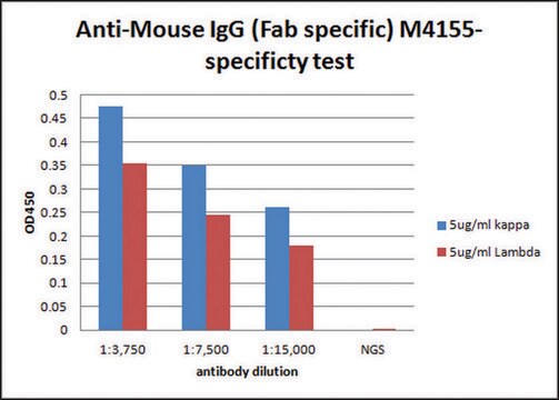
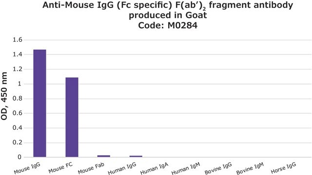
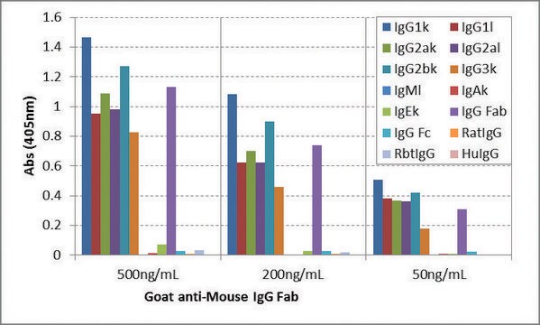
Anti-Mouse IgG (Fab specific) F(ab′)2 fragment antibody is monospecific as determined by immunoelectrophoresis versus normal mouse serum, mouse IgG and the Fab fragment of mouse IgG. The antibody does not react with the Fc fragment of mouse IgG. Moreover, no reactivity is observed with human κ or lambda light chain, IgG, IgA, IgM, IgD and IgE, bovine IgG and IgM, or horse IgG as determined by ELISA. In immunoelectrophoresis, the antibody preparation is found to consist only of the F(ab′)2 fragment of goat IgG, where specific antisera is applied to goat IgG fragments. No contamination with goat IgG whole molecule is observed.
Anti-Mouse IgG (Fab specific) F(ab′)2 fragment antibody is monospecific as determined by immunoelectrophoresis versus normal mouse serum, mouse IgG and the Fab fragment of mouse IgG. The antibody does not react with the Fc fragment of mouse IgG. Moreover, no reactivity is observed with human κ or lambda light chain, IgG, IgA, IgM, IgD and IgE, bovine IgG and IgM, or horse IgG as determined by ELISA. In immunoelectrophoresis, the antibody preparation is found to consist only of the F(ab′)2 fragment of goat IgG, where specific antisera is applied to goat IgG fragments. No contamination with goat IgG whole molecule is observed.
Immunogen
Mouse IgG
Application
Anti-Mouse IgG (Fab specific) F(ab′)2 fragment antibody is suitable for use in ELISA (using a conc. Of 2μg/mI (100 μg/well) in 50 mM carbonate/bicarbonate buffer, at pH9.6) . The antibody can also be used in immunoelectrophoresis.
Other Notes
Antibody adsorbed with bovine, equine and human serum proteins.
Physical form
Solution in 0.01 M phosphate buffered saline, pH 7.4, containing 15 mM sodium azide.
Disclaimer
Unless otherwise stated in our catalog or other company documentation accompanying the product(s), our products are intended for research use only and are not to be used for any other purpose, which includes but is not limited to, unauthorized commercial uses, in vitro diagnostic uses, ex vivo or in vivo therapeutic uses or any type of consumption or application to humans or animals.
Not finding the right product?
Try our Product Selector Tool.
Storage Class Code
12 - Non Combustible Liquids
WGK
nwg
Flash Point(F)
Not applicable
Flash Point(C)
Not applicable
Choose from one of the most recent versions:
Already Own This Product?
Find documentation for the products that you have recently purchased in the Document Library.
Customers Also Viewed
T E Steinsvik et al.
Scandinavian journal of immunology, 42(6), 607-616 (1995-12-01)
SCID and RAG-2 deficient mice were transplanted intraperitoneally with human peripheral blood lymphocytes (hu-PBL-SCID and hu-PBL-RAG mice). Seven days after transplantation the mice were immunized with a pneumococcal polysaccharide vaccine. Flow cytometry analysis of cells from the peritoneal cavity and
The Ocular Conjunctiva as a Mucosal Immunization Route: A Profile of the Immune Response to the Model Antigen Tetanus Toxoid
Barisani-Asenbauer T, et al.
PLoS ONE, 8(4) (2013)
Peter Engelhardt et al.
Methods in molecular biology (Clifton, N.J.), 369, 387-405 (2007-07-28)
Standard immunogold-labeling methods in transmission electron microscopy (TEM) are unable to locate immunogold particles in the depth direction. This inability does not only concern bulky whole mounts, but also sections. A partial solution to the problem is stereo inspection. However
J Wang et al.
Journal of neuroscience research, 50(1), 23-31 (1997-10-23)
The role of the transcription factor AP-1 in regulating D2 receptor transcriptional activity was investigated in D2 receptor expressing neuroblastoma cells, NB41A3, and in non-D2 receptor expressing CHO cells. Deletion of a region containing the putative AP-1 binding site resulted
Chen Yang et al.
Molecular medicine reports, 18(2), 1353-1360 (2018-06-15)
Previous studies have demonstrated that lipid rafts and β‑adducin serve an important role in leukocyte rolling. In the present study the migratory ability and behavior of neutrophils was demonstrated to rely on the integrity of the lipid raft structure. β‑adducin
Our team of scientists has experience in all areas of research including Life Science, Material Science, Chemical Synthesis, Chromatography, Analytical and many others.
Contact Technical Service