Skip to Content
Merck
All Photos(3)

Key Documents

ABS204

Sigma-Aldrich

Anti-phospho-PDHE1-A type I (Ser293) Antibody

from rabbit, purified by affinity chromatography

Synonym(s):

Pyruvate dehydrogenase E1 component subunit alpha, somatic form, mitochondrial, PDHE1-A type I

Sign Into View Organizational & Contract Pricing


About This Item

UNSPSC Code:
12352203
eCl@ss:
32160702
NACRES:
NA.41

biological source

rabbit

Quality Level

antibody form

affinity isolated antibody

antibody product type

primary antibodies

clone

polyclonal

purified by

affinity chromatography

species reactivity

human

species reactivity (predicted by homology)

primate (based on 100% sequence homology), zebrafish (based on 100% sequence homology), rat (based on 100% sequence homology), bovine (based on 100% sequence homology), mouse (based on 100% sequence homology)

technique(s)

immunocytochemistry: suitable
immunoprecipitation (IP): suitable
western blot: suitable

NCBI accession no.

UniProt accession no.

shipped in

wet ice

target post-translational modification

phosphorylation (pSer293)

Gene Information

human ... PDHA1(5160)

General description

In many organisms, the pyruvate dehydrogenase (PDH) complex catalyzes the overall, irreversible conversion of pyruvate to acetyl-CoA and CO2 in the aerobic pathway. This complex also serves as a key regulator for cardiac substrate selection. It contains multiple copies of three enzymatic components: pyruvate dehydrogenase (E1 or PDHE1-A type I), dihydrolipoamide acetyltransferase (E2), and lipoamide dehydrogenase (E3). PDH is regulated by both pyruvate dehydrogenase kinase (PDK)-mediated phosphorylation and feedback inhibition.

Specificity

This antibody recognizes PDHE1-A type I phosphorylated at Ser293.

Immunogen

Epitope: Phosphorylated Ser293
KLH-conjugated linear peptide corresponding to human PDHE1-A type I phosphorylated at Ser293.

Application

Detect phospho-PDHE1-A type I (Ser293) using this Anti-phospho-PDHE1-A type I (Ser293) Antibody validated for use in WB, IC & IP.
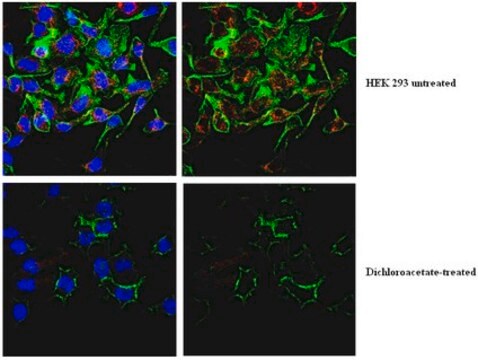
Immunocytochemistry Analysis: A 1:500 dilution of this antibody from a representative lot detected phospho-PDHE1-A type I (Ser293) in untreated and DCA-treated HEK293 cells.
Research Category
Metabolism
Research Sub Category
Glucose/Glycogen Metabolism

Quality

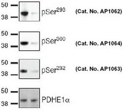
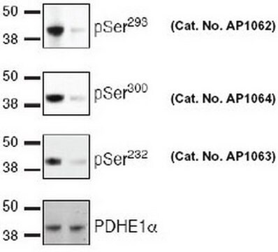
Evaluated by Western Blot in DCA untreated and treated HEK293 cell lysates.

Western Blot Analysis: A 1:10,000 dilution of this antibody detected phospho-PDHE1-A type I (Ser293) in untreated and DCA-treated HEK293 cell lysates.

Target description

~43 kDa observed

Physical form

Affinity purified
Purified rabbit polyclonal in buffer containing 0.1 M Tris-Glycine (pH 7.4), 150 mM NaCl with 0.05% sodium azide.

Storage and Stability

Stable for 1 year at 2-8°C from date of receipt.

Analysis Note

Control
Untreated and DCA-treated HEK293 cell lysates

Disclaimer

Unless otherwise stated in our catalog or other company documentation accompanying the product(s), our products are intended for research use only and are not to be used for any other purpose, which includes but is not limited to, unauthorized commercial uses, in vitro diagnostic uses, ex vivo or in vivo therapeutic uses or any type of consumption or application to humans or animals.

Not finding the right product?  

Try our Product Selector Tool.

Storage Class Code

12 - Non Combustible Liquids

WGK

WGK 1

Flash Point(F)

Not applicable

Flash Point(C)

Not applicable


Certificates of Analysis (COA)

Search for Certificates of Analysis (COA) by entering the products Lot/Batch Number. Lot and Batch Numbers can be found on a product’s label following the words ‘Lot’ or ‘Batch’.

Already Own This Product?

Find documentation for the products that you have recently purchased in the Document Library.

Visit the Document Library

Ian M Willis et al.
Proceedings of the National Academy of Sciences of the United States of America, 115(48), 12182-12187 (2018-11-16)
As a master negative regulator of RNA polymerase (Pol) III, Maf1 modulates transcription in response to nutrients and stress to balance the production of highly abundant tRNAs, 5S rRNA, and other small noncoding RNAs with cell growth and maintenance. This
Ratnakar Tiwari et al.
iScience, 25(10), 105086-105086 (2022-09-27)
Endothelial cell (EC) metabolism has emerged as a driver of angiogenesis. While hypoxia inactivates the oxygen sensors prolyl-4 hydroxylase domain-containing proteins 1-3 (PHD1-3) and stimulates angiogenesis, the effects of PHDs on EC functions remain poorly defined. Here, we investigated the
Cyril Corbet et al.
Nature communications, 9(1), 1208-1208 (2018-03-25)
Lactate exchange between glycolytic and oxidative cancer cells is proposed to optimize tumor growth. Blocking lactate uptake through monocarboxylate transporter 1 (MCT1) represents an attractive therapeutic strategy but may stimulate glucose consumption by oxidative cancer cells. We report here that
Hui Wang et al.
EMBO reports, 22(9), e52247-e52247 (2021-08-07)
Our knowledge of the coordination of fuel usage in skeletal muscle is incomplete. Whether and how microRNAs are involved in the substrate selection for oxidation is largely unknown. Here we show that mice lacking miR-183 and miR-96 have enhanced muscle
Stephen W Standage et al.
American journal of physiology. Heart and circulatory physiology, 312(2), H239-H249 (2016-11-25)
Children with sepsis and multisystem organ failure have downregulated leukocyte gene expression of peroxisome proliferator-activated receptor-α (PPARα), a nuclear hormone receptor transcription factor that regulates inflammation and lipid metabolism. Mouse models of sepsis have likewise demonstrated that the absence of

Our team of scientists has experience in all areas of research including Life Science, Material Science, Chemical Synthesis, Chromatography, Analytical and many others.

Contact Technical Service