Skip to Content
Merck
All Photos(1)

Documents

201M-9

Sigma-Aldrich

Actin, Muscle Specific (HHF35) Mouse Monoclonal Antibody

Sign Into View Organizational & Contract Pricing


About This Item

UNSPSC Code:
12352203
NACRES:
NA.41

biological source

mouse

Quality Level

100
500

conjugate

unconjugated

antibody form

culture supernatant

antibody product type

primary antibodies

clone

HHF35, monoclonal

description

For In Vitro Diagnostic Use in Select Regions (See Chart)

form

buffered aqueous solution

species reactivity

human

packaging

vial of 0.1 mL concentrate (201M-94)
vial of 0.5 mL concentrate (201M-95)
bottle of 1.0 mL predilute (201M-97)
vial of 1.0 mL concentrate (201M-96)
bottle of 7.0 mL predilute (201M-98)

manufacturer/tradename

Cell Marque

technique(s)

immunohistochemistry (formalin-fixed, paraffin-embedded sections): 1:10-1:50

isotype

IgG1κ

control

skeletal muscle

shipped in

wet ice

storage temp.

2-8°C

visualization

cytoplasmic

General description

Mucins are a diverse group of complex, highly glycosylated extracellular proteins. Mucin 2 (MUC2) protects the gastric and intestinal epithelium from chemical and mechanical injury. Anti-MUC2 reactivity is normally seen in goblet cells of the small intestine and colon and is associated with mucinous carcinomas, including those of the gastrointestinal tract and ovary. MUC2 immunohistochemistry is useful for identifying colonic, gastric, and esophageal carcinomas.
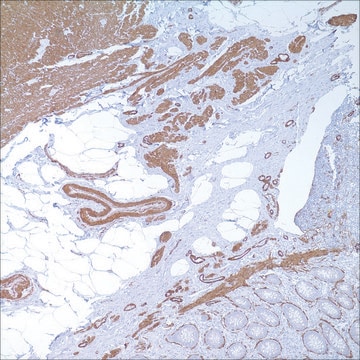
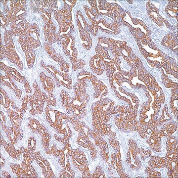
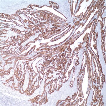
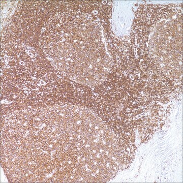
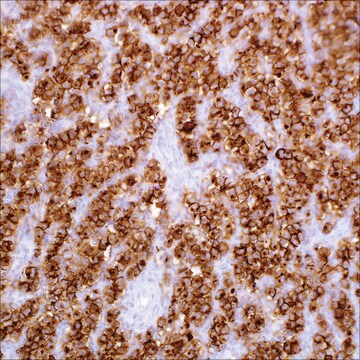
Muscle Specific Actin is a part of the actin family of proteins which are highly conserved, major components of the cytoskeleton. Anti-Muscle Specific Actin immunohistochemical reactivity is seen in skeletal, cardiac, and smooth muscle cells and can be seen in neoplasms with muscle differentiation such as leiomyomas and rhabdomyosarcomas. In contrast, anti-Muscle Specific Actin reactivity is typically not seen in endothelial cells, connective tissues, carcinomas, melanomas, lymphomas, and most non-myogenic sarcomas.

Quality


IVD

IVD

IVD

RUO

Linkage

Actin, Muscle Specific Positive Control Slides, Product No. 201S, are available for immunohistochemistry (formalin-fixed, paraffin-embedded sections).

Physical form

Solution in Tris Buffer, pH 7.3-7.7, with 1% BSA and <0.1% Sodium Azide

Preparation Note

Download the IFU specific to your product lot and formatNote: This requires a keycode which can be found on your packaging or product label.

Other Notes

For Technical Service please contact: 800-665-7284 or email: service@cellmarque.com

Legal Information

Cell Marque is a trademark of Merck KGaA, Darmstadt, Germany

Not finding the right product?  

Try our Product Selector Tool.


Certificates of Analysis (COA)

Search for Certificates of Analysis (COA) by entering the products Lot/Batch Number. Lot and Batch Numbers can be found on a product’s label following the words ‘Lot’ or ‘Batch’.

Already Own This Product?

Find documentation for the products that you have recently purchased in the Document Library.

Visit the Document Library

R A Schmidt et al.
The American journal of pathology, 131(1), 19-28 (1988-04-01)
The authors have recently developed a monoclonal antibody, HHF35, that recognizes the muscle-specific isoforms of actin. To determine its potential usefulness in the differential diagnosis of "small, round, blue cell" tumors of childhood, they immunolabeled formalinor B-5-fixed tissue sections from
N Azumi et al.
Modern pathology : an official journal of the United States and Canadian Academy of Pathology, Inc, 1(6), 469-474 (1988-11-01)
Two fibrillary proteins, muscle-specific actin (MSA) and desmin, are found only in cells of smooth and skeletal muscle lineages. Among the monoclonal antibodies (MAbs) to these antigens which we have tested, we found several to be reactive in formalin-fixed, paraffin-embedded
S Rangdaeng et al.
American journal of clinical pathology, 96(1), 32-45 (1991-07-01)
Muscle-specific actin (MSA) and desmin are considered to be sensitive and specific markers for muscle differentiation. The authors compared staining patterns for these markers in 576 samples of normal, reactive, and neoplastic tissues. The standard avidin-biotin-peroxidase complex technique was performed
T Tsukada et al.
The American journal of pathology, 127(2), 389-402 (1987-05-01)
Monoclonal antibody HHF35 has previously been characterized biochemically as recognizing isotypes of actin (alpha and gamma) which are specific to muscle cells. In this study, the authors have investigated the normal and pathologic tissue distribution of HHF35-positive cells using the
A M Gown et al.
The American journal of pathology, 125(1), 191-207 (1986-10-01)
The authors have performed immunocytochemical investigations of the distribution of various cell types in human atherosclerotic plaques using monoclonal antibodies specific to smooth muscle cells (CGA7 [Gown et al, J Cell Biol 1985, 100:807-813] and HHF35 [Tsukada et al, Am

Our team of scientists has experience in all areas of research including Life Science, Material Science, Chemical Synthesis, Chromatography, Analytical and many others.

Contact Technical Service