SCT264
BioTracker™ Mitophagy Live Cell Probe
Synonym(s):
mitochondrial degradation probe, mitophagy detection, mitophagy probe
Sign Into View Organizational & Contract Pricing
All Photos(3)
About This Item
UNSPSC Code:
41106514
Recommended Products
biological source
human
Quality Level
form
lyophilized solid
packaging
vial of 15 μg
manufacturer/tradename
Millipore
technique(s)
cell culture | stem cell: suitable
shipped in
2-8°C
storage temp.
2-8°C
General description
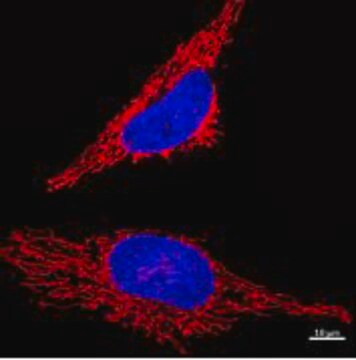
The Mitophagy probe accumulates in intact mitochondria, where it is immobilized by chemical bonds and exhibits a weak fluorescence under normal conditions. When mitophagy is induced, the damaged mitochondria fuse with the lysosome, increasing fluorescence
SPECTRAL PROPERTIES
λex: 530 nm
λem: 700 nm
A 100 uM stock solution of the mitophagy probe is created by dissolving 50 uL DMSO to one tube (5 ug) of mitophagy dye.
A typical working solution concentration is 1 nM.
SPECTRAL PROPERTIES
λex: 530 nm
λem: 700 nm
A 100 uM stock solution of the mitophagy probe is created by dissolving 50 uL DMSO to one tube (5 ug) of mitophagy dye.
A typical working solution concentration is 1 nM.
Features and Benefits
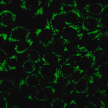
SCT264 live cell mitophagy detection probe accumulates and is immobilized on intact mitochondria, which fuse to the lysosome after mitophagy is induced, causing the probe to fluoresce brightly. Mitophagy has been associated with the accumulation of dysfunctional mitochondria in diseases such Alzheimer′s and Parkinson′s.
Target description
Mitochondria play an essential role in cells for the production of energy. Mitophagy, or the removal of damaged mitochondria via autophagy, has been connected to Alzheimer s and Parkinson s disease induced by the accumulation of depolarized mitochondria. Mitophagy serves as a dedicated elimination system that removes mitochondria whose dysfunction may be caused by oxidative stress and DNA damage. Mitochondria destined for autophagy are sequestered into the autophagosome, fused to the lysosome, and digested.
Physical form
purple solid
Storage and Stability
Store at 2 - 8 °C, protected from light.
Other Notes
This far red live cell probe for mitophagy detection fluoresces brightly when damaged mitochondria enter the acidic environment of the lysosome.
Legal Information
BioTracker is a trademark of Merck KGaA, Darmstadt, Germany
Disclaimer
Unless otherwise stated in our catalog or other company documentation accompanying the product(s), our products are intended for research use only and are not to be used for any other purpose, which includes but is not limited to, unauthorized commercial uses, in vitro diagnostic uses, ex vivo or in vivo therapeutic uses or any type of consumption or application to humans or animals.
Storage Class Code
11 - Combustible Solids
WGK
WGK 3
Flash Point(F)
Not applicable
Flash Point(C)
Not applicable
Certificates of Analysis (COA)
Search for Certificates of Analysis (COA) by entering the products Lot/Batch Number. Lot and Batch Numbers can be found on a product’s label following the words ‘Lot’ or ‘Batch’.
Already Own This Product?
Find documentation for the products that you have recently purchased in the Document Library.
Our team of scientists has experience in all areas of research including Life Science, Material Science, Chemical Synthesis, Chromatography, Analytical and many others.
Contact Technical Service