Saltar al contenido
Merck
Todas las fotos(1)

Key Documents

SAB5300247

Sigma-Aldrich

Monoclonal Anti-MMP9 antibody produced in mouse

clone 5G3, ascites fluid

Sinónimos:

CLG4B, GELB, MANDP2, MMP-9

Iniciar sesiónpara Ver la Fijación de precios por contrato y de la organización


About This Item

UNSPSC Code:
12352203
NACRES:
NA.41

biological source

mouse

conjugate

unconjugated

antibody form

ascites fluid

antibody product type

primary antibodies

clone

5G3, monoclonal

mol wt

92 kDa

species reactivity

human

technique(s)

direct ELISA: 1:10,000
flow cytometry: 1:200-1:400
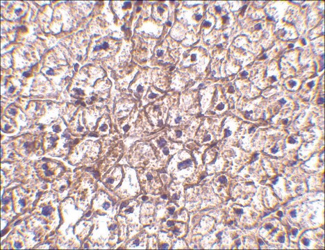
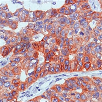
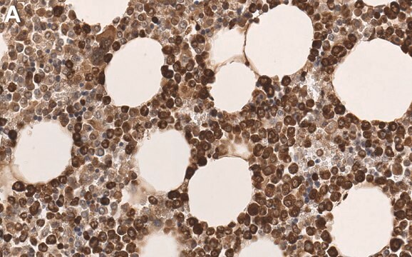
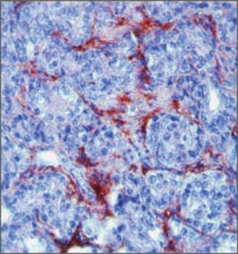
immunohistochemistry: 1:200-1:1,000
indirect immunofluorescence: 1:200-1:1,000
western blot: 1:500-1:2,000

isotype

IgG2a

NCBI accession no.

UniProt accession no.

shipped in

wet ice

storage temp.

−20°C

target post-translational modification

unmodified

Gene Information

human ... MMP9(4318)

Immunogen

Purified recombinant fragment of human MMP9 expressed in E.coli.
Mouse monoclonal antibody raised against MMP9

Physical form

Ascitic fluid containing 0.03% sodium azide.

Disclaimer

Unless otherwise stated in our catalog or other company documentation accompanying the product(s), our products are intended for research use only and are not to be used for any other purpose, which includes but is not limited to, unauthorized commercial uses, in vitro diagnostic uses, ex vivo or in vivo therapeutic uses or any type of consumption or application to humans or animals.

¿No encuentra el producto adecuado?  

Pruebe nuestro Herramienta de selección de productos.

Storage Class

10 - Combustible liquids

wgk_germany

WGK 3

flash_point_f

Not applicable

flash_point_c

Not applicable


Certificados de análisis (COA)

Busque Certificados de análisis (COA) introduciendo el número de lote del producto. Los números de lote se encuentran en la etiqueta del producto después de las palabras «Lot» o «Batch»

¿Ya tiene este producto?

Encuentre la documentación para los productos que ha comprado recientemente en la Biblioteca de documentos.

Visite la Librería de documentos

Qi Gao et al.
Oncotarget, 8(55), 93899-93911 (2017-12-08)
Although materials and modern surgical techniques have been developed to suppress postoperative adhesions, adhesion formation can still occur, and thus, a novel effective anti-adhesion drug is greatly needed. In the present study, we explored the efficacy of paeoniflorin treatment against
Zhengjiang Li et al.
Experimental and therapeutic medicine, 24(6), 750-750 (2022-12-24)
Proline- and serine-rich 2 (PROSER2) is encoded by the 47th open reading frame on human chromosome 10. Bioinformatic analysis has shown PROSER2 was significantly correlated with prognostic outcome of osteosarcoma patients. Its role in the progression and metastasis of human
Heiani Touaitahuata et al.
Developmental biology, 393(1), 57-70 (2014-07-06)
During long bone development and post-natal growth, the cartilaginous model of the skeleton is progressively replaced by bone, a process known as endochondral ossification. In the primary spongiosa, osteoclasts degrade the mineralized cartilage produced by hypertrophic chondrocytes to generate cartilage
Spencer Slavin et al.
Carcinogenesis, 35(6), 1301-1309 (2014-01-01)
The prostate cancer (PCa) microenvironment contains active stromal cells known as cancer-associated fibroblasts (CAF) that may play important roles in influencing tumor progression. Here we studied the role of CAF estrogen receptor alpha (ERα) and found that it could protect
Jingyin Chen et al.
Journal of pineal research, 57(3), 340-347 (2014-09-05)
Melatonin is a strong anti-oxidant that has beneficial effects against early brain injury (EBI) following a subarachnoid hemorrhage (SAH) in rats; protection includes the reduction of both mortality and neurological deficits. The molecular mechanisms underlying these clinical effects in the

Nuestro equipo de científicos tiene experiencia en todas las áreas de investigación: Ciencias de la vida, Ciencia de los materiales, Síntesis química, Cromatografía, Analítica y muchas otras.

Póngase en contacto con el Servicio técnico